8JIY | pdb_00008jiy

A carbohydrate binding domain of a putative chondroitinase


Experimental Data Snapshot

  • Method: X-RAY DIFFRACTION
  • Resolution: 1.55 Å
  • R-Value Free: 
    0.228 (Depositor), 0.229 (DCC) 
  • R-Value Work: 
    0.191 (Depositor), 0.191 (DCC) 
  • R-Value Observed: 
    0.193 (Depositor) 

Starting Model: in silico
View more details

wwPDB Validation 3D Report Full Report

Validation slider image for 8JIY

This is version 1.1 of the entry. See complete history

Literature

Identification and structural characterization of a novel chondroitin sulfate-specific carbohydrate-binding module: The first member of a new family, CBM100.

Liu, G.Chang, Y.Mei, X.Chen, G.Zhang, Y.Jiang, X.Tao, W.Xue, C.

(2024) Int J Biol Macromol 255: 127959-127959

  • DOI: https://doi.org/10.1016/j.ijbiomac.2023.127959
  • Primary Citation Related Structures: 
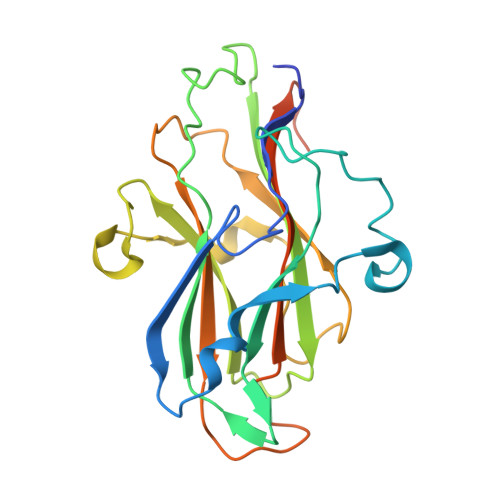
    8JIY

  • PubMed Abstract: 

    Chondroitin sulfate is a biologically and commercially important polysaccharide with a variety of applications. Carbohydrate-binding module (CBM) is an important class of carbohydrate-binding protein, which could be utilized as a promising tool for the applications of polysaccharides. In the present study, an unknown function domain was explored from a putative chondroitin sulfate lyase in PL29 family. Recombinant PhCBM100 demonstrated binding capacity to chondroitin sulfates with K a values of 2.1 ± 0.2 × 10 6  M -1 and 6.0 ± 0.1 × 10 6  M -1 to chondroitin sulfate A and chondroitin sulfate C, respectively. The 1.55 Å resolution X-ray crystal structure of PhCBM100 exhibited a β-sandwich fold formed by two antiparallel β-sheets. A binding groove in PhCBM100 interacting with chondroitin sulfate was subsequently identified, and the potential of PhCBM100 for visualization of chondroitin sulfate was evaluated. PhCBM100 is the first characterized chondroitin sulfate-specific CBM. The novelty of PhCBM100 proposed a new CBM family of CBM100.


  • Organizational Affiliation
    • College of Food Science and Engineering, Ocean University of China, 1299 Sansha Road, Qingdao 266404, China.

Macromolecule Content 

  • Total Structure Weight: 27.12 kDa 
  • Atom Count: 1,976 
  • Modeled Residue Count: 216 
  • Deposited Residue Count: 241 
  • Unique protein chains: 1

Macromolecules

Find similar proteins by:|  3D Structure
Entity ID: 1
MoleculeChains  Sequence LengthOrganismDetailsImage
DUF4955 domain-containing protein241Polaribacter haliotisMutation(s): 0 
Gene Names: H9I45_07735
UniProt
Find proteins for A0A7L8AJZ3 (Polaribacter haliotis)
Explore A0A7L8AJZ3 
Go to UniProtKB:  A0A7L8AJZ3
Entity Groups
Sequence Clusters30% Identity50% Identity70% Identity90% Identity95% Identity100% Identity
UniProt GroupA0A7L8AJZ3
Sequence Annotations
Expand
Reference Sequence

Experimental Data & Validation

Experimental Data

  • Method: X-RAY DIFFRACTION
  • Resolution: 1.55 Å
  • R-Value Free:  0.228 (Depositor), 0.229 (DCC) 
  • R-Value Work:  0.191 (Depositor), 0.191 (DCC) 
  • R-Value Observed: 0.193 (Depositor) 
Space Group: P 21 21 21
Unit Cell:
Length ( Å )Angle ( ˚ )
a = 56.313α = 90
b = 56.634β = 90
c = 69.456γ = 90
Software Package:
Software NamePurpose
PHENIXrefinement
Aimlessdata scaling
XDSdata reduction
PDB_EXTRACTdata extraction
PHASERphasing

Structure Validation

View Full Validation Report


Entry History 

& Funding Information

Deposition Data


Funding OrganizationLocationGrant Number
Not funded--

Revision History  (Full details and data files)

  • Version 1.0: 2023-09-20
    Type: Initial release
  • Version 1.1: 2024-04-03
    Changes: Database references