8JBK | pdb_00008jbk

Crystal structure of Na+,K+-ATPase in the E1.3Na+ state


Experimental Data Snapshot

  • Method: X-RAY DIFFRACTION
  • Resolution: 2.80 Å
  • R-Value Free: 
    0.270 (Depositor), 0.274 (DCC) 
  • R-Value Work: 
    0.229 (Depositor), 0.235 (DCC) 
  • R-Value Observed: 
    0.231 (Depositor) 

Starting Model: experimental
View more details

wwPDB Validation 3D Report Full Report

Validation slider image for 8JBK

This is version 1.2 of the entry. See complete history

Literature

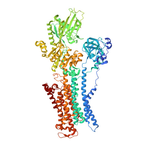
Crystal structures of Na + ,K + -ATPase reveal the mechanism that converts the K + -bound form to Na + -bound form and opens and closes the cytoplasmic gate.

Kanai, R.Vilsen, B.Cornelius, F.Toyoshima, C.

(2023) FEBS Lett 597: 1957-1976

  • DOI: https://doi.org/10.1002/1873-3468.14689
  • Primary Citation Related Structures: 
    8JBK, 8JBL, 8JBM, 8JFZ

  • PubMed Abstract: 

    Na + ,K + -ATPase (NKA) plays a pivotal role in establishing electrochemical gradients for Na + and K + across the cell membrane by alternating between the E1 (showing high affinity for Na + and low affinity for K + ) and E2 (low affinity to Na + and high affinity to K + ) forms. Presented here are two crystal structures of NKA in E1·Mg 2+ and E1·3Na + states at 2.9 and 2.8 Å resolution, respectively. These two E1 structures fill a gap in our description of the NKA reaction cycle based on the atomic structures. We describe how NKA converts the K + -bound E2·2K + form to an E1 (E1·Mg 2+ ) form, which allows high-affinity Na + binding, eventually closing the cytoplasmic gate (in E1 ~ P·ADP·3Na + ) after binding three Na + , while keeping the extracellular ion pathway sealed. We now understand previously unknown functional roles for several parts of NKA and that NKA uses even the lipid bilayer for gating the ion pathway.


  • Organizational Affiliation
    • Institute for Quantitative Biosciences, The University of Tokyo, Bunkyo-ku, Japan.

Macromolecule Content 

  • Total Structure Weight: 330.25 kDa 
  • Atom Count: 21,682 
  • Modeled Residue Count: 2,641 
  • Deposited Residue Count: 2,778 
  • Unique protein chains: 3

Macromolecules

Find similar proteins by:|  3D Structure
Entity ID: 1
MoleculeChains  Sequence LengthOrganismDetailsImage
Sodium/potassium-transporting ATPase subunit alphaA,
D [auth C]
1,021Sus scrofaMutation(s): 0 
EC: 7.2.2.13
Membrane Entity: Yes 
UniProt
Find proteins for P05024 (Sus scrofa)
Explore P05024 
Go to UniProtKB:  P05024
Entity Groups
Sequence Clusters30% Identity50% Identity70% Identity90% Identity95% Identity100% Identity
UniProt GroupP05024
Sequence Annotations
Expand
Reference Sequence
Find similar proteins by:|  3D Structure
Entity ID: 2
MoleculeChains  Sequence LengthOrganismDetailsImage
Sodium/potassium-transporting ATPase subunit beta-1B,
E [auth D]
303Sus scrofaMutation(s): 0 
Membrane Entity: Yes 
UniProt
Find proteins for P05027 (Sus scrofa)
Explore P05027 
Go to UniProtKB:  P05027
Entity Groups
Sequence Clusters30% Identity50% Identity70% Identity90% Identity95% Identity100% Identity
UniProt GroupP05027
Glycosylation
Glycosylation Sites: 2
Sequence Annotations
Expand
Reference Sequence
Find similar proteins by:|  3D Structure
Entity ID: 3
MoleculeChains  Sequence LengthOrganismDetailsImage
FXYD domain-containing ion transport regulatorC [auth G],
F [auth E]
65Sus scrofaMutation(s): 0 
Membrane Entity: Yes 
UniProt
Find proteins for Q58K79 (Sus scrofa)
Explore Q58K79 
Go to UniProtKB:  Q58K79
Entity Groups
Sequence Clusters30% Identity50% Identity70% Identity90% Identity95% Identity100% Identity
UniProt GroupQ58K79
Sequence Annotations
Expand
Reference Sequence

Oligosaccharides

Help  
Entity ID: 4
MoleculeChains Length2D Diagram GlycosylationD Interactions
alpha-D-mannopyranose-(1-6)-alpha-D-mannopyranose-(1-6)-[alpha-D-mannopyranose-(1-3)]beta-D-mannopyranose-(1-4)-2-acetamido-2-deoxy-beta-D-glucopyranose-(1-4)-2-acetamido-2-deoxy-beta-D-glucopyranoseG [auth F]6N-Glycosylation
Glycosylation Resources
GlyTouCan: G34442SS
GlyCosmos: G34442SS
GlyGen: G34442SS
Entity ID: 5
MoleculeChains Length2D Diagram GlycosylationD Interactions
alpha-D-mannopyranose-(1-3)-[alpha-D-mannopyranose-(1-6)]beta-D-mannopyranose-(1-4)-2-acetamido-2-deoxy-beta-D-glucopyranose-(1-4)-2-acetamido-2-deoxy-beta-D-glucopyranose
H
5N-Glycosylation
Glycosylation Resources
GlyTouCan: G22768VO
GlyCosmos: G22768VO
GlyGen: G22768VO
Entity ID: 6
MoleculeChains Length2D Diagram GlycosylationD Interactions
2-acetamido-2-deoxy-beta-D-glucopyranose-(1-4)-2-acetamido-2-deoxy-beta-D-glucopyranose
I
2N-Glycosylation
Glycosylation Resources
GlyTouCan: G42666HT
GlyCosmos: G42666HT
GlyGen: G42666HT

Small Molecules

Ligands 7 Unique
IDChains Name / Formula / InChI Key2D Diagram3D Interactions
PC1

Query on PC1


Download:Ideal Coordinates CCD File
Q [auth A],
S [auth A],
T [auth A]
1,2-DIACYL-SN-GLYCERO-3-PHOSPHOCHOLINE
C44 H88 N O8 P
NRJAVPSFFCBXDT-HUESYALOSA-N
PCW

Query on PCW


Download:Ideal Coordinates CCD File
HA [auth C]
IA [auth C]
JA [auth C]
KA [auth C]
LA [auth C]
HA [auth C],
IA [auth C],
JA [auth C],
KA [auth C],
LA [auth C],
MA [auth C],
NA [auth C],
OA [auth C],
R [auth A],
U [auth A],
V [auth A],
W [auth A],
X [auth A],
Y [auth A],
Z [auth B]
1,2-DIOLEOYL-SN-GLYCERO-3-PHOSPHOCHOLINE
C44 H85 N O8 P
SNKAWJBJQDLSFF-NVKMUCNASA-O
DMU

Query on DMU


Download:Ideal Coordinates CCD File
SA [auth E]DECYL-BETA-D-MALTOPYRANOSIDE
C22 H42 O11
WOQQAWHSKSSAGF-WXFJLFHKSA-N
CLR

Query on CLR


Download:Ideal Coordinates CCD File
AA [auth B]
GA [auth C]
O [auth A]
P [auth A]
PA [auth D]
AA [auth B],
GA [auth C],
O [auth A],
P [auth A],
PA [auth D],
RA [auth E]
CHOLESTEROL
C27 H46 O
HVYWMOMLDIMFJA-DPAQBDIFSA-N
NAG

Query on NAG


Download:Ideal Coordinates CCD File
QA [auth D]2-acetamido-2-deoxy-beta-D-glucopyranose
C8 H15 N O6
OVRNDRQMDRJTHS-FMDGEEDCSA-N
MN

Query on MN


Download:Ideal Coordinates CCD File
BA [auth C],
J [auth A]
MANGANESE (II) ION
Mn
WAEMQWOKJMHJLA-UHFFFAOYSA-N
NA
(Subject of Investigation/LOI)

Query on NA


Download:Ideal Coordinates CCD File
CA [auth C]
DA [auth C]
EA [auth C]
FA [auth C]
K [auth A]
CA [auth C],
DA [auth C],
EA [auth C],
FA [auth C],
K [auth A],
L [auth A],
M [auth A],
N [auth A]
SODIUM ION
Na
FKNQFGJONOIPTF-UHFFFAOYSA-N

Experimental Data & Validation

Experimental Data

  • Method: X-RAY DIFFRACTION
  • Resolution: 2.80 Å
  • R-Value Free:  0.270 (Depositor), 0.274 (DCC) 
  • R-Value Work:  0.229 (Depositor), 0.235 (DCC) 
  • R-Value Observed: 0.231 (Depositor) 
Space Group: P 1 21 1
Unit Cell:
Length ( Å )Angle ( ˚ )
a = 193.74α = 90
b = 74.49β = 115.72
c = 162.56γ = 90
Software Package:
Software NamePurpose
BSSdata collection
HKL-2000data reduction
STARANISOdata scaling
PHASERphasing
PHENIXrefinement

Structure Validation

View Full Validation Report


Entry History 

& Funding Information

Deposition Data


Funding OrganizationLocationGrant Number
Japan Society for the Promotion of Science (JSPS)JapanJP19H00975
Japan Society for the Promotion of Science (JSPS)JapanJP21K06109

Revision History  (Full details and data files)

  • Version 1.0: 2023-08-09
    Type: Initial release
  • Version 1.1: 2023-08-16
    Changes: Data collection, Database references
  • Version 1.2: 2024-11-13
    Changes: Structure summary