8JA6 | pdb_00008ja6

Structure of the alginate epimerase/lyase complexed with tri-mannuronic acid


Experimental Data Snapshot

  • Method: X-RAY DIFFRACTION
  • Resolution: 2.00 Å
  • R-Value Free: 
    0.178 (Depositor), 0.178 (DCC) 
  • R-Value Work: 
    0.154 (Depositor), 0.155 (DCC) 
  • R-Value Observed: 
    0.155 (Depositor) 

Starting Model: experimental
View more details

wwPDB Validation 3D Report Full Report

Validation slider image for 8JA6

This is version 1.2 of the entry. See complete history

Literature

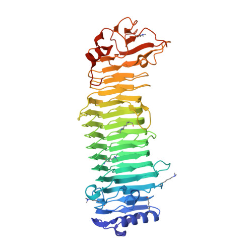
Structural basis for the minimal bifunctional alginate epimerase AlgE3 from Azotobacter chroococcum.

Fujiwara, T.Mano, E.Nango, E.

(2024) FEBS Lett 598: 1422-1437

  • DOI: https://doi.org/10.1002/1873-3468.14886
  • Primary Citation Related Structures: 
    8JA4, 8JA6, 8JAZ, 8XFQ, 8XFR

  • PubMed Abstract: 

    Among the epimerases specific to alginate, some of them in Azotobacter genera convert β-d-mannuronic acid to α-l-guluronic acid but also have lyase activity to degrade alginate. The remarkable characteristics of these epimerases make it a promising enzyme for tailoring alginates to meet specific demands. Here, we determined the structure of the bifunctional mannuronan C-5 epimerase AlgE3 from Azotobacter chroococcum (AcAlgE3) in complex with several mannuronic acid oligomers as well as in apo form, which allowed us to elucidate the binding manner of each mannuronic acid oligomer, and the structural plasticity, which is dependent on calcium ions. Moreover, a comprehensive analysis of the lyase activity profiles of AcAlgE3 combined with structural characteristics explained the preference for different chain length oligomers.


  • Organizational Affiliation
    • Institute of Multidisciplinary Research for Advanced Materials, Tohoku University, Sendai, Japan.

Macromolecule Content 

  • Total Structure Weight: 53.69 kDa 
  • Atom Count: 3,887 
  • Modeled Residue Count: 480 
  • Deposited Residue Count: 493 
  • Unique protein chains: 1

Macromolecules

Find similar proteins by:|  3D Structure
Entity ID: 1
MoleculeChains  Sequence LengthOrganismDetailsImage
mannuronan 5-epimerase493Azotobacter chroococcum NCIMB 8003Mutation(s): 0 
Gene Names: algE7Achr_39570
EC: 5.1.3.37
UniProt
Find proteins for A0A0C4WKK2 (Azotobacter chroococcum NCIMB 8003)
Explore A0A0C4WKK2 
Go to UniProtKB:  A0A0C4WKK2
Entity Groups
Sequence Clusters30% Identity50% Identity70% Identity90% Identity95% Identity100% Identity
UniProt GroupA0A0C4WKK2
Sequence Annotations
Expand
Reference Sequence

Oligosaccharides

Help  
Entity ID: 2
MoleculeChains Length2D Diagram GlycosylationD Interactions
beta-D-mannopyranuronic acid-(1-4)-beta-D-mannopyranuronic acid-(1-4)-beta-D-mannopyranuronic acid
B
3N/A
Glycosylation Resources
GlyTouCan: G85027VZ
GlyCosmos: G85027VZ

Small Molecules

Modified Residues  1 Unique
IDChains TypeFormula2D DiagramParent
MLY
Query on MLY
A
L-PEPTIDE LINKINGC8 H18 N2 O2LYS

Experimental Data & Validation

Experimental Data

  • Method: X-RAY DIFFRACTION
  • Resolution: 2.00 Å
  • R-Value Free:  0.178 (Depositor), 0.178 (DCC) 
  • R-Value Work:  0.154 (Depositor), 0.155 (DCC) 
  • R-Value Observed: 0.155 (Depositor) 
Space Group: H 3
Unit Cell:
Length ( Å )Angle ( ˚ )
a = 121.71α = 90
b = 121.71β = 90
c = 120.69γ = 120
Software Package:
Software NamePurpose
XDSdata reduction
XSCALEdata scaling
PHASERphasing
PHENIXrefinement

Structure Validation

View Full Validation Report


Entry History 

& Funding Information

Deposition Data

  • Released Date: 2024-05-08 
  • Deposition Author(s): Fujiwara, T.

Funding OrganizationLocationGrant Number
Japan Society for the Promotion of Science (JSPS)Japan21K06077

Revision History  (Full details and data files)

  • Version 1.0: 2024-05-08
    Type: Initial release
  • Version 1.1: 2024-05-15
    Changes: Database references
  • Version 1.2: 2024-06-26
    Changes: Database references