8J9T | pdb_00008j9t

Crystal Structure of GyraseA N-terminal at 2.43A Resolution


Experimental Data Snapshot

  • Method: X-RAY DIFFRACTION
  • Resolution: 2.43 Å
  • R-Value Free: 
    0.235 (Depositor), 0.240 (DCC) 
  • R-Value Work: 
    0.199 (Depositor), 0.200 (DCC) 

Starting Model: experimental
View more details

wwPDB Validation 3D Report Full Report

Validation slider image for 8J9T

This is version 1.1 of the entry. See complete history

Literature

Structural and Comparative Stability of a Truncated N-Terminal Domain of DNA Gyrase A From Salmonella Typhi.

Salman, M.Sachdeva, E.Negi, S.Das, U.Ethayathulla, A.S.Kaur, P.

(2025) Proteins 

  • DOI: https://doi.org/10.1002/prot.70070
  • Primary Citation Related Structures: 
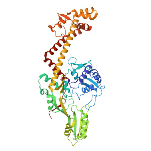
    8J9T

  • PubMed Abstract: 

    DNA Gyrase, a Type II topoisomerase, introduces negative supercoiling in dsDNA through the cleavage and religation activity at the expense of ATP. DNA Gyrase forms a hetero-tetrameric complex with two Gyrase A and Gyrase B subunits. These two subunits interact dynamically to physically transfer one DNA duplex through another by coupling ATP binding and hydrolysis with DNA binding, cleavage, and strand transport. The N-terminal domain of Gyrase A (GyrA-NTD) mediates the cleavage of the DNA strand and forms the target site for quinolones class of antibiotics. While structures of GyrA-NTD from several prokaryotes have been determined, the N-terminal segment (residues 1-32) remains unresolved in apo forms. Here, we present the crystal structure of a truncated GyrA-NTD (ΔGyrA-NTD; residues 33-530) from Salmonella Typhi at 2.43 Å resolution, alongside comparative biophysical characterization with the wild type. Thermal and chemical denaturation assays revealed that the wild-type GyrA-NTD is more prone to unfolding than the truncated variant, indicating that deletion of the unresolved N-terminal segment enhances domain stability. These findings uncover a structural element influencing GyrA-NTD stability.


  • Organizational Affiliation
    • All India Institute of Medical Sciences, New Delhi Department of Biophysics, Delhi, India.

Macromolecule Content 

  • Total Structure Weight: 110.87 kDa 
  • Atom Count: 8,337 
  • Modeled Residue Count: 974 
  • Deposited Residue Count: 986 
  • Unique protein chains: 1

Macromolecules

Find similar proteins by:|  3D Structure
Entity ID: 1
MoleculeChains  Sequence LengthOrganismDetailsImage
DNA gyrase subunit A
A, B
493Salmonella enterica subsp. enterica serovar Typhi str. CT18Mutation(s): 0 
Gene Names: gyrA
EC: 5.6.2.2
UniProt
Find proteins for A0A716CHN5 (Salmonella enterica subsp. enterica serovar Typhi str. CT18)
Explore A0A716CHN5 
Go to UniProtKB:  A0A716CHN5
Entity Groups
Sequence Clusters30% Identity50% Identity70% Identity90% Identity95% Identity100% Identity
UniProt GroupA0A716CHN5
Sequence Annotations
Expand
Reference Sequence

Experimental Data & Validation

Experimental Data

  • Method: X-RAY DIFFRACTION
  • Resolution: 2.43 Å
  • R-Value Free:  0.235 (Depositor), 0.240 (DCC) 
  • R-Value Work:  0.199 (Depositor), 0.200 (DCC) 
Space Group: P 61
Unit Cell:
Length ( Å )Angle ( ˚ )
a = 154.781α = 90
b = 154.781β = 90
c = 108.258γ = 120
Software Package:
Software NamePurpose
REFMACrefinement
XDSdata reduction
Aimlessdata scaling
MOLREPphasing
MxCuBEdata collection
Cootmodel building

Structure Validation

View Full Validation Report


Entry History 

& Funding Information

Deposition Data


Funding OrganizationLocationGrant Number
Not funded--

Revision History  (Full details and data files)

  • Version 1.0: 2024-05-08
    Type: Initial release
  • Version 1.1: 2025-11-26
    Changes: Database references, Structure summary