8J9K | pdb_00008j9k

Structure of basal beta-arrestin2


Experimental Data Snapshot

  • Method: ELECTRON MICROSCOPY
  • Resolution: 3.50 Å
  • Aggregation State: PARTICLE 
  • Reconstruction Method: SINGLE PARTICLE 

Starting Model: experimental
View more details

wwPDB Validation 3D Report Full Report

Validation slider image for 8J9K

This is version 1.2 of the entry. See complete history

Literature

Molecular insights into atypical modes of beta-arrestin interaction with seven transmembrane receptors.

Maharana, J.Sano, F.K.Sarma, P.Yadav, M.K.Duan, L.Stepniewski, T.M.Chaturvedi, M.Ranjan, A.Singh, V.Saha, S.Mahajan, G.Chami, M.Shihoya, W.Selent, J.Chung, K.Y.Banerjee, R.Nureki, O.Shukla, A.K.

(2024) Science 383: 101-108

  • DOI: https://doi.org/10.1126/science.adj3347
  • Primary Citation Related Structures: 
    8GO9, 8J8R, 8J8V, 8J8Z, 8J97, 8J9K, 8JA3, 8JAF

  • PubMed Abstract: 

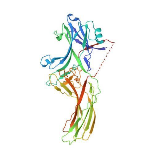
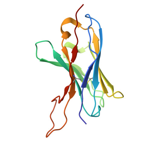
    β-arrestins (βarrs) are multifunctional proteins involved in signaling and regulation of seven transmembrane receptors (7TMRs), and their interaction is driven primarily by agonist-induced receptor activation and phosphorylation. Here, we present seven cryo-electron microscopy structures of βarrs either in the basal state, activated by the muscarinic receptor subtype 2 (M2R) through its third intracellular loop, or activated by the βarr-biased decoy D6 receptor (D6R). Combined with biochemical, cellular, and biophysical experiments, these structural snapshots allow the visualization of atypical engagement of βarrs with 7TMRs and also reveal a structural transition in the carboxyl terminus of βarr2 from a β strand to an α helix upon activation by D6R. Our study provides previously unanticipated molecular insights into the structural and functional diversity encoded in 7TMR-βarr complexes with direct implications for exploring novel therapeutic avenues.


  • Organizational Affiliation
    • Department of Biological Sciences, Indian Institute of Technology Kanpur, Kanpur, India.

Macromolecule Content 

  • Total Structure Weight: 69.48 kDa 
  • Atom Count: 4,284 
  • Modeled Residue Count: 590 
  • Deposited Residue Count: 624 
  • Unique protein chains: 3

Macromolecules

Find similar proteins by:|  3D Structure
Entity ID: 1
MoleculeChains  Sequence LengthOrganismDetailsImage
Beta-arrestin-2A [auth C]393Rattus norvegicusMutation(s): 0 
Gene Names: Arrb2
UniProt
Find proteins for P29067 (Rattus norvegicus)
Explore P29067 
Go to UniProtKB:  P29067
Entity Groups
Sequence Clusters30% Identity50% Identity70% Identity90% Identity95% Identity100% Identity
UniProt GroupP29067
Sequence Annotations
Expand
Reference Sequence
Find similar proteins by:|  3D Structure
Entity ID: 2
MoleculeChains  Sequence LengthOrganismDetailsImage
Fab6 light chain106Mus musculusMutation(s): 0 
Find similar proteins by:|  3D Structure
Entity ID: 3
MoleculeChains  Sequence LengthOrganismDetailsImage
Fab6 heavy chainC [auth D]125Mus musculusMutation(s): 0 

Experimental Data & Validation

Experimental Data

  • Method: ELECTRON MICROSCOPY
  • Resolution: 3.50 Å
  • Aggregation State: PARTICLE 
  • Reconstruction Method: SINGLE PARTICLE 
EM Software:
TaskSoftware PackageVersion
MODEL REFINEMENTPHENIX
RECONSTRUCTIONcryoSPARC3.3.1

Structure Validation

View Full Validation Report


Entry History 

& Funding Information

Deposition Data


Funding OrganizationLocationGrant Number
Science and Engineering Research Board (SERB)IndiaIPA/2020/000405
Department of Biotechnology (DBT, India)IndiaIA/S/20/1/504916
Science and Engineering Research Board (SERB)IndiaCRG/2022/002646
Science and Engineering Research Board (SERB)IndiaSPR/2020/000408

Revision History  (Full details and data files)

  • Version 1.0: 2023-12-27
    Type: Initial release
  • Version 1.1: 2024-01-17
    Changes: Database references
  • Version 1.2: 2024-11-20
    Changes: Data collection, Structure summary