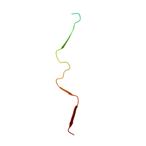
E22G A beta 40 fibril structure and kinetics illuminate how A beta 40 rather than A beta 42 triggers familial Alzheimer's.
Tehrani, M.J., Matsuda, I., Yamagata, A., Kodama, Y., Matsunaga, T., Sato, M., Toyooka, K., McElheny, D., Kobayashi, N., Shirouzu, M., Ishii, Y.(2024) Nat Commun 15: 7045-7045
- PubMed: 39147751
- DOI: https://doi.org/10.1038/s41467-024-51294-w
- Primary Citation of Related Structures:
8J47 - PubMed Abstract:
Arctic (E22G) mutation in amyloid-β (Aβ enhances Aβ40 fibril accumulation in Alzheimer's disease (AD). Unlike sporadic AD, familial AD (FAD) patients with the mutation exhibit more Aβ40 in the plaque core. However, structural details of E22G Aβ40 fibrils remain elusive, hindering therapeutic progress. Here, we determine a distinctive W-shaped parallel β-sheet structure through co-analysis by cryo-electron microscopy (cryoEM) and solid-state nuclear magnetic resonance (SSNMR) of in-vitro-prepared E22G Aβ40 fibrils. The E22G Aβ40 fibrils displays typical amyloid features in cotton-wool plaques in the FAD, such as low thioflavin-T fluorescence and a less compact unbundled morphology. Furthermore, kinetic and MD studies reveal previously unidentified in-vitro evidence that E22G Aβ40, rather than Aβ42, may trigger Aβ misfolding in the FAD, and prompt subsequent misfolding of wild-type (WT) Aβ40/Aβ42 via cross-seeding. The results provide insight into how the Arctic mutation promotes AD via Aβ40 accumulation and cross-propagation.
Organizational Affiliation:
School of Life Science and Technology, Tokyo Institute of Technology, 4259 Nagatsuta-cho, Midori-ku, Yokohama, Kanagawa, 226-8503, Japan.


