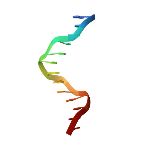
8J0K

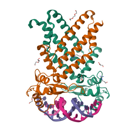
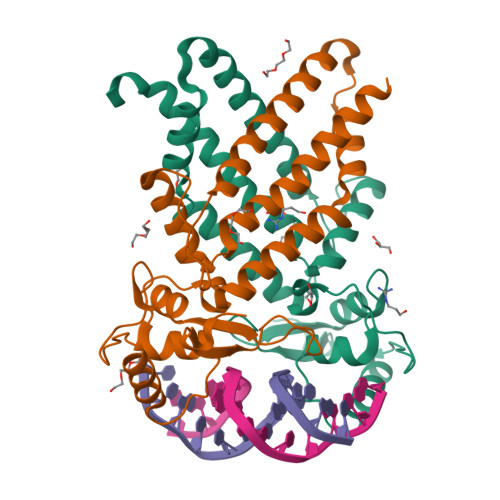
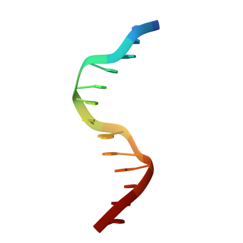
Crystal structure of human TFAP2A in complex with DNA


Experimental Data Snapshot

  • Method: X-RAY DIFFRACTION
  • Resolution: 2.10 Å
  • R-Value Free: 0.196 
  • R-Value Work: 0.165 
  • R-Value Observed: 0.166 

Starting Model: experimental
View more details

wwPDB Validation   3D Report Full Report


This is version 1.2 of the entry. See complete history


Literature

Structural basis for specific DNA sequence motif recognition by the TFAP2 transcription factors.

Liu, K.Xiao, Y.Gan, L.Li, W.Zhang, J.Min, J.

(2023) Nucleic Acids Res 51: 8270-8282

  • DOI: https://doi.org/10.1093/nar/gkad583
  • Primary Citation of Related Structures:  
    8J0K, 8J0L, 8J0Q, 8J0R

  • PubMed Abstract: 

    The TFAP2 family regulates gene expression during differentiation, development, and organogenesis, and includes five homologs in humans. They all possess a highly conserved DNA binding domain (DBD) followed by a helix-span-helix (HSH) domain. The DBD-HSH tandem domain specifically binds to a GCC(N3)GGC consensus sequence, but the precise recognition mechanisms remain unclear. Here, we found that TFAP2 preferred binding to the GCC(N3)GGC sequence, and the pseudo-palindromic GCC and GGC motifs and the length of the central spacer between the two motifs determined their binding specificity. Structural studies revealed that the two flat amphipathic α-helical HSH domains of TFAP2A stacked with each other to form a dimer via hydrophobic interactions, while the stabilized loops from both DBD domains inserted into two neighboring major grooves of the DNA duplex to form base-specific interactions. This specific DNA binding mechanism controlled the length of the central spacer and determined the DNA sequence specificity of TFAP2. Mutations of the TFAP2 proteins are implicated in various diseases. We illustrated that reduction or disruption of the DNA binding ability of the TFAP2 proteins is the primary cause of TFAP2 mutation-associated diseases. Thus, our findings also offer valuable insights into the pathogenesis of disease-associated mutations in TFAP2 proteins.


  • Organizational Affiliation

    Hubei Key Laboratory of Genetic Regulation and Integrative Biology, School of Life Sciences, Central China Normal University, Wuhan 430079, PR China.


Macromolecules

Find similar proteins by:  (by identity cutoff)  |  3D Structure
Entity ID: 1
MoleculeChains Sequence LengthOrganismDetailsImage
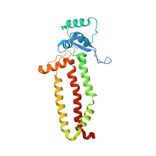
Transcription factor AP-2-alpha
A, B
219Homo sapiensMutation(s): 0 
Gene Names: TFAP2AAP2TFTFAP2
UniProt & NIH Common Fund Data Resources
Find proteins for P05549 (Homo sapiens)
Explore P05549 
Go to UniProtKB:  P05549
PHAROS:  P05549
GTEx:  ENSG00000137203 
Entity Groups  
Sequence Clusters30% Identity50% Identity70% Identity90% Identity95% Identity100% Identity
UniProt GroupP05549
Sequence Annotations
Expand
  • Reference Sequence
Small Molecules
Ligands 3 Unique
IDChains Name / Formula / InChI Key2D Diagram3D Interactions
PGE
Query on PGE

Download Ideal Coordinates CCD File 
E [auth A]
F [auth A]
I [auth B]
J [auth B]
L [auth B]
TRIETHYLENE GLYCOL
C6 H14 O4
ZIBGPFATKBEMQZ-UHFFFAOYSA-N
PG3
Query on PG3

Download Ideal Coordinates CCD File 
H [auth A],
K [auth B]
GUANIDINE-3-PROPANOL
C4 H12 N3 O
JDXXTKLHHZMVIO-UHFFFAOYSA-O
GOL
Query on GOL

Download Ideal Coordinates CCD File 
G [auth A],
N [auth B]
GLYCEROL
C3 H8 O3
PEDCQBHIVMGVHV-UHFFFAOYSA-N
Experimental Data & Validation

Experimental Data

  • Method: X-RAY DIFFRACTION
  • Resolution: 2.10 Å
  • R-Value Free: 0.196 
  • R-Value Work: 0.165 
  • R-Value Observed: 0.166 
  • Space Group: P 32 2 1
Unit Cell:
Length ( Å )Angle ( ˚ )
a = 124.261α = 90
b = 124.261β = 90
c = 96.926γ = 120
Software Package:
Software NamePurpose
REFMACrefinement
XDSdata reduction
autoPROCdata scaling
PHASERphasing

Structure Validation

View Full Validation Report


Entry History & Funding Information

Deposition Data


Funding OrganizationLocationGrant Number
National Natural Science Foundation of China (NSFC)China31770834

Revision History  (Full details and data files)

  • Version 1.0: 2023-07-05
    Type: Initial release
  • Version 1.1: 2023-08-30
    Changes: Data collection, Database references
  • Version 1.2: 2023-09-06
    Changes: Database references