8IUC | pdb_00008iuc

Crystal structure of GH65 alpha-1,2-glucosidase from Flavobacterium johnsoniae in complex with isomaltose


Experimental Data Snapshot

  • Method: X-RAY DIFFRACTION
  • Resolution: 1.56 Å
  • R-Value Free: 
    0.211 (Depositor), 0.217 (DCC) 
  • R-Value Work: 
    0.187 (Depositor), 0.193 (DCC) 

Starting Model: experimental
View more details

wwPDB Validation 3D Report Full Report

Validation slider image for 8IUC

This is version 1.2 of the entry. See complete history

Literature

Bacteroidota polysaccharide utilization system for branched dextran exopolysaccharides from lactic acid bacteria.

Nakamura, S.Kurata, R.Tonozuka, T.Funane, K.Park, E.Y.Miyazaki, T.

(2023) J Biological Chem 299: 104885-104885

  • DOI: https://doi.org/10.1016/j.jbc.2023.104885
  • Primary Citation Related Structures: 
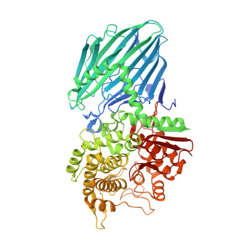
    8IU8, 8IU9, 8IUA, 8IUB, 8IUC

  • PubMed Abstract: 

    Dextran is an α-(1→6)-glucan that is synthesized by some lactic acid bacteria, and branched dextran with α-(1→2)-, α-(1→3)-, and α-(1→4)-linkages are often produced. Although many dextranases are known to act on the α-(1→6)-linkage of dextran, few studies have functionally analyzed the proteins involved in degrading branched dextran. The mechanism by which bacteria utilize branched dextran is unknown. Earlier, we identified dextranase (FjDex31A) and kojibiose hydrolase (FjGH65A) in the dextran utilization locus (FjDexUL) of a soil Bacteroidota Flavobacterium johnsoniae and hypothesized that FjDexUL is involved in the degradation of α-(1→2)-branched dextran. In this study, we demonstrate that FjDexUL proteins recognize and degrade α-(1→2)- and α-(1→3)-branched dextrans produced by Leuconostoc citreum S-32 (S-32 α-glucan). The FjDexUL genes were significantly upregulated when S-32 α-glucan was the carbon source compared with α-glucooligosaccharides and α-glucans, such as linear dextran and branched α-glucan from L. citreum S-64. FjDexUL glycoside hydrolases synergistically degraded S-32 α-glucan. The crystal structure of FjGH66 shows that some sugar-binding subsites can accommodate α-(1→2)- and α-(1→3)-branches. The structure of FjGH65A in complex with isomaltose supports that FjGH65A acts on α-(1→2)-glucosyl isomaltooligosaccharides. Furthermore, two cell surface sugar-binding proteins (FjDusD and FjDusE) were characterized, and FjDusD showed an affinity for isomaltooligosaccharides and FjDusE for dextran, including linear and branched dextrans. Collectively, FjDexUL proteins are suggested to be involved in the degradation of α-(1→2)- and α-(1→3)-branched dextrans. Our results will be helpful in understanding the bacterial nutrient requirements and symbiotic relationships between bacteria at the molecular level.


  • Organizational Affiliation
    • Department of Bioscience, Graduate School of Science and Technology, Shizuoka University, Suruga-ku, Shizuoka, Japan.

Macromolecule Content 

  • Total Structure Weight: 232.36 kDa 
  • Atom Count: 17,596 
  • Modeled Residue Count: 1,977 
  • Deposited Residue Count: 2,034 
  • Unique protein chains: 1

Macromolecules

Find similar proteins by:|  3D Structure
Entity ID: 1
MoleculeChains  Sequence LengthOrganismDetailsImage
Candidate alpha glycoside phosphorylase Glycoside hydrolase family 65
A, B, C
678Flavobacterium johnsoniae UW101Mutation(s): 0 
Gene Names: Fjoh_4428
EC: 3.2.1.216
UniProt
Find proteins for A5FBJ5 (Flavobacterium johnsoniae (strain ATCC 17061 / DSM 2064 / JCM 8514 / BCRC 14874 / CCUG 350202 / NBRC 14942 / NCIMB 11054 / UW101))
Explore A5FBJ5 
Go to UniProtKB:  A5FBJ5
Entity Groups
Sequence Clusters30% Identity50% Identity70% Identity90% Identity95% Identity100% Identity
UniProt GroupA5FBJ5
Sequence Annotations
Expand
Reference Sequence

Oligosaccharides

Help  
Entity ID: 2
MoleculeChains Length2D Diagram GlycosylationD Interactions
alpha-D-glucopyranose-(1-6)-beta-D-glucopyranose
D, E, F
2N/A
Glycosylation Resources
GlyTouCan: G45624LJ
GlyCosmos: G45624LJ

Small Molecules

Ligands 1 Unique
IDChains Name / Formula / InChI Key2D Diagram3D Interactions
EDO

Query on EDO


Download:Ideal Coordinates CCD File
AA [auth C]
BA [auth C]
CA [auth C]
DA [auth C]
EA [auth C]
AA [auth C],
BA [auth C],
CA [auth C],
DA [auth C],
EA [auth C],
FA [auth C],
G [auth A],
GA [auth C],
H [auth A],
HA [auth C],
I [auth A],
IA [auth C],
J [auth A],
JA [auth C],
K [auth A],
L [auth A],
M [auth A],
N [auth A],
O [auth A],
P [auth A],
Q [auth B],
R [auth B],
S [auth B],
T [auth B],
U [auth B],
V [auth B],
W [auth B],
X [auth B],
Y [auth B],
Z [auth B]
1,2-ETHANEDIOL
C2 H6 O2
LYCAIKOWRPUZTN-UHFFFAOYSA-N

Experimental Data & Validation

Experimental Data

  • Method: X-RAY DIFFRACTION
  • Resolution: 1.56 Å
  • R-Value Free:  0.211 (Depositor), 0.217 (DCC) 
  • R-Value Work:  0.187 (Depositor), 0.193 (DCC) 
Space Group: C 1 2 1
Unit Cell:
Length ( Å )Angle ( ˚ )
a = 122.938α = 90
b = 194.09β = 116.348
c = 111.88γ = 90
Software Package:
Software NamePurpose
REFMACrefinement
XDSdata reduction
Aimlessdata scaling
MOLREPphasing

Structure Validation

View Full Validation Report


Entry History 

& Funding Information

Deposition Data


Funding OrganizationLocationGrant Number
Japan Society for the Promotion of Science (JSPS)Japan19K15748

Revision History  (Full details and data files)

  • Version 1.0: 2023-06-14
    Type: Initial release
  • Version 1.1: 2023-07-12
    Changes: Database references
  • Version 1.2: 2024-05-29
    Changes: Data collection