8IM0 | pdb_00008im0

mCherry-LaM8 complex


Experimental Data Snapshot

  • Method: X-RAY DIFFRACTION
  • Resolution: 1.31 Å
  • R-Value Free: 
    0.190 (Depositor), 0.192 (DCC) 
  • R-Value Work: 
    0.178 (Depositor), 0.178 (DCC) 
  • R-Value Observed: 
    0.178 (Depositor) 

Starting Model: experimental
View more details

wwPDB Validation 3D Report Full Report

Validation slider image for 8IM0

This is version 2.0 of the entry. See complete history

Literature

Structural Insights into the Binding of Red Fluorescent Protein mCherry-Specific Nanobodies.

Liang, H.Ma, Z.Wang, Z.Zhong, P.Li, R.Jiang, H.Zong, X.Zhong, C.Liu, X.Liu, P.Liu, J.Zhu, H.Liu, R.Ding, Y.

(2023) Int J Mol Sci 24

  • DOI: https://doi.org/10.3390/ijms24086952
  • Primary Citation Related Structures: 
    8ILX, 8IM0, 8IM1

  • PubMed Abstract: 

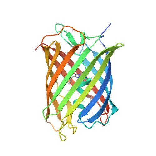
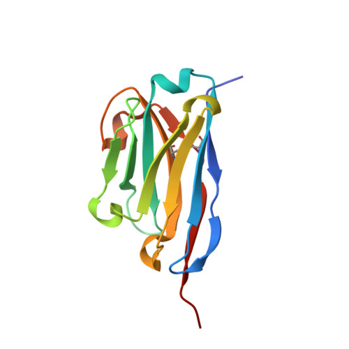
    Red fluorescent proteins (RFPs) have broad applications in life science research, and the manipulation of RFPs using nanobodies can expand their potential uses. However, the structural information available for nanobodies that bind with RFPs is still insufficient. In this study, we cloned, expressed, purified, and crystallized complexes formed by mCherry with LaM1, LaM3, and LaM8. Then, we analyzed the biochemical properties of the complexes using mass spectrometry (MS), fluorescence-detected size exclusion chromatography (FSEC), isothermal titration calorimetry (ITC), and bio-layer interferometry (BLI) technology. We determined the crystal structure of mCherry-LaM1, mCherry-LaM3, and mCherry-LaM8, with resolutions of 2.05 Å, 3.29 Å, and 1.31 Å, respectively. In this study, we systematically compared various parameters of several LaM series nanobodies, including LaM1, LaM3, and LaM8, with previously reported data on LaM2, LaM4, and LaM6, specifically examining their structural information. After designing multivalent tandem LaM1-LaM8 and LaM8-LaM4 nanobodies based on structural information, we characterized their properties, revealing their higher affinity and specificity to mCherry. Our research provides novel structural insights that could aid in understanding nanobodies targeting a specific target protein. This could provide a starting point for developing enhanced mCherry manipulation tools.


  • Organizational Affiliation
    • State Key Laboratory of Genetic Engineering, School of Life Sciences, Fudan University, Shanghai 200438, China.

Macromolecule Content 

  • Total Structure Weight: 40.62 kDa 
  • Atom Count: 3,231 
  • Modeled Residue Count: 339 
  • Deposited Residue Count: 362 
  • Unique protein chains: 2

Macromolecules

Find similar proteins by:|  3D Structure
Entity ID: 1
MoleculeChains  Sequence LengthOrganismDetailsImage
MCherry fluorescent protein235Anaplasma marginaleMutation(s): 1 
Gene Names: mCherry
Find similar proteins by:|  3D Structure
Entity ID: 2
MoleculeChains  Sequence LengthOrganismDetailsImage
LaM8127Camelus bactrianusMutation(s): 0 

Small Molecules

Modified Residues  1 Unique
IDChains TypeFormula2D DiagramParent
CH6
Query on CH6
A
L-PEPTIDE LINKINGC16 H19 N3 O4 SMET, TYR, GLY

Experimental Data & Validation

Experimental Data

  • Method: X-RAY DIFFRACTION
  • Resolution: 1.31 Å
  • R-Value Free:  0.190 (Depositor), 0.192 (DCC) 
  • R-Value Work:  0.178 (Depositor), 0.178 (DCC) 
  • R-Value Observed: 0.178 (Depositor) 
Space Group: P 21 21 21
Unit Cell:
Length ( Å )Angle ( ˚ )
a = 63.075α = 90
b = 75.31β = 90
c = 83.625γ = 90
Software Package:
Software NamePurpose
PHENIXrefinement
Aimlessdata scaling
DIALSdata reduction
PHASERphasing

Structure Validation

View Full Validation Report


Entry History 

& Funding Information

Deposition Data


Funding OrganizationLocationGrant Number
National Science Foundation (NSF, China)China32070939
National Science Foundation (NSF, China)China82030106

Revision History  (Full details and data files)

  • Version 1.0: 2023-06-21
    Type: Initial release
  • Version 1.1: 2024-11-13
    Changes: Data collection, Structure summary
  • Version 2.0: 2026-03-18
    Changes: Polymer sequence