8II8

X-ray crystal structure of pink-colored protein from Pleurotus salmoneostramineus in complex with natural chromophore


Experimental Data Snapshot

  • Method: X-RAY DIFFRACTION
  • Resolution: 1.25 Å
  • R-Value Free: 0.164 
  • R-Value Work: 0.131 

Starting Model: experimental
View more details

wwPDB Validation   3D Report Full Report


Ligand Structure Quality Assessment 


This is version 1.1 of the entry. See complete history


Literature

Crystal Structure of the Native Chromoprotein from Pleurotus salmoneostramineus Provides Insights into the Pigmentation Mechanism.

Ihara, M.Tsuchida, N.Sumida, M.Himiyama, T.Kitayama, T.Shirasaka, N.Fukuta, Y.

(2024) J Agric Food Chem 72: 17626-17632

  • DOI: https://doi.org/10.1021/acs.jafc.4c02951
  • Primary Citation of Related Structures:  
    8II8

  • PubMed Abstract: 

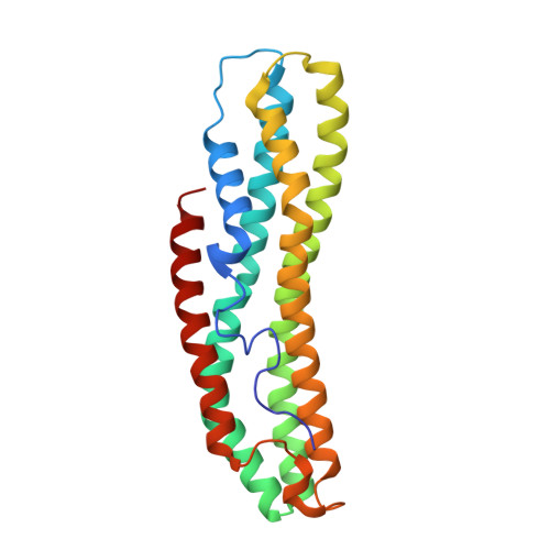
    The pink-colored protein from the fungus Pleurotus salmoneostramineus (PsPCP) possesses unusual primary sequences with little resemblance to those of known proteins and exhibits a red color in aqueous solution. To understand the pigmentation mechanism of PsPCP, we elucidated the X-ray crystal structure of the native PsPCP. We identified a highly conjugated polyene ligand 2-dehydro-3-deoxylaetiporic acid A as a chromophore ligand, whose solution exhibits yellow. The crystal structure of PsPCP indicated that the ligand is secured in the central cavity and anchored at both termini by hydrophilic interactions and that surrounding residues show CH-pi and C-H···O hydrogen bondings. Geometrical analyses of the bound ligand demonstrated that the conjugated C-C and C═C bonds exhibit similar bond distances. The result indicated enhanced electron delocalization within the conjugated CC bond system, resulting in a redshift of the chromophore ligand. The computational estimates of the UV-vis spectra support the view that the electron delocalization within the conjugated CC bonds system of the bound ligand, induced by the specific ligand geometry within a limited space of PsPCP cavity, is responsible for the red pigmentation of PsPCP. Thus, we propose that the coloring mechanism of PsPCP, which constrains the geometry of a highly conjugated polyene ligand, is a novel type of pigment chemistry.


  • Organizational Affiliation

    Faculty of Agriculture, Kindai University, 3327-204 Nakamachi, Nara, Nara 631-8505, Japan.


Macromolecules
Find similar proteins by:  (by identity cutoff)  |  3D Structure
Entity ID: 1
MoleculeChains Sequence LengthOrganismDetailsImage
Chromoprotein226Pleurotus salmoneostramineusMutation(s): 0 
UniProt
Find proteins for A0A2Z5U5S2 (Pleurotus salmoneostramineus)
Explore A0A2Z5U5S2 
Go to UniProtKB:  A0A2Z5U5S2
Entity Groups  
Sequence Clusters30% Identity50% Identity70% Identity90% Identity95% Identity100% Identity
UniProt GroupA0A2Z5U5S2
Sequence Annotations
Expand
  • Reference Sequence
Small Molecules
Ligands 2 Unique
IDChains Name / Formula / InChI Key2D Diagram3D Interactions
PW6 (Subject of Investigation/LOI)
Query on PW6

Download Ideal Coordinates CCD File 
C [auth A](2E,5E,7Z,9E,11E,13E,15Z,17E,19Z,21E,23E)-24-methyl-25-oxohexacosa-2,5,7,9,11,13,15,17,19,21,23-undecaenoic acid
C27 H30 O3
ZUWHINCAQDXMJU-JXLQPSFDSA-N
ACE
Query on ACE

Download Ideal Coordinates CCD File 
B [auth A]ACETYL GROUP
C2 H4 O
IKHGUXGNUITLKF-UHFFFAOYSA-N
Experimental Data & Validation

Experimental Data

  • Method: X-RAY DIFFRACTION
  • Resolution: 1.25 Å
  • R-Value Free: 0.164 
  • R-Value Work: 0.131 
  • Space Group: I 1 2 1
Unit Cell:
Length ( Å )Angle ( ˚ )
a = 58.879α = 90
b = 31.895β = 95.939
c = 107.416γ = 90
Software Package:
Software NamePurpose
REFMACrefinement
XDSdata reduction
Aimlessdata scaling
MOLREPphasing

Structure Validation

View Full Validation Report


Ligand Structure Quality Assessment 


Entry History & Funding Information

Deposition Data


Funding OrganizationLocationGrant Number
Japan Society for the Promotion of Science (JSPS)Japan19K06175

Revision History  (Full details and data files)

  • Version 1.0: 2024-08-14
    Type: Initial release
  • Version 1.1: 2024-08-21
    Changes: Database references