8I28 | pdb_00008i28

Structure of Phosphoserine Aminotransferase from Saccharomyces cerevisiae


Experimental Data Snapshot

  • Method: X-RAY DIFFRACTION
  • Resolution: 2.80 Å
  • R-Value Free: 
    0.268 (Depositor), 0.264 (DCC) 
  • R-Value Work: 
    0.229 (Depositor), 0.230 (DCC) 
  • R-Value Observed: 
    0.231 (Depositor) 

Starting Model: experimental
View more details

wwPDB Validation 3D Report Full Report

Validation slider image for 8I28

This is version 1.1 of the entry. See complete history

Literature

Molecular Structure of Phosphoserine Aminotransferase from Saccharomyces cerevisiae.

Jang, J.Chang, J.H.

(2023) Int J Mol Sci 24

  • DOI: https://doi.org/10.3390/ijms24065139
  • Primary Citation Related Structures: 
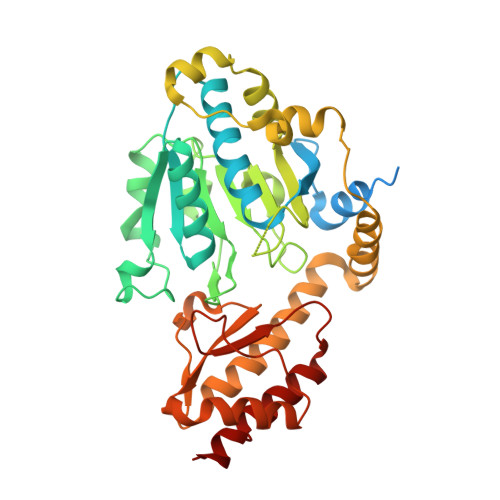
    8I28

  • PubMed Abstract: 

    Phosphoserine aminotransferase (PSAT) is a pyridoxal 5'-phosphate-dependent enzyme involved in the second step of the phosphorylated pathway of serine biosynthesis. PSAT catalyzes the transamination of 3-phosphohydroxypyruvate to 3-phosphoserine using L-glutamate as the amino donor. Although structural studies of PSAT have been performed from archaea and humans, no structural information is available from fungi. Therefore, to elucidate the structural features of fungal PSAT, we determined the crystal structure of Saccharomyces cerevisiae PSAT ( Sc PSAT) at a resolution of 2.8 Å. The results demonstrated that the Sc PSAT protein was dimeric in its crystal structure. Moreover, the gate-keeping loop of Sc PSAT exhibited a conformation similar to that of other species. Several distinct structural features in the halide-binding and active sites of Sc PSAT were compared with its homologs. Overall, this study contributes to our current understanding of PSAT by identifying the structural features of fungal PSAT for the first time.


  • Organizational Affiliation
    • Department of Biology Education, Kyungpook National University, 80 Daehak-ro, Buk-gu, Daegu 41566, Republic of Korea.

Macromolecule Content 

  • Total Structure Weight: 86.94 kDa 
  • Atom Count: 5,253 
  • Modeled Residue Count: 670 
  • Deposited Residue Count: 790 
  • Unique protein chains: 1

Macromolecules

Find similar proteins by:|  3D Structure
Entity ID: 1
MoleculeChains  Sequence LengthOrganismDetailsImage
Phosphoserine aminotransferase
A, B
395Saccharomyces cerevisiaeMutation(s): 0 
Gene Names: SER1SERCYOR184W
EC: 2.6.1.52
UniProt
Find proteins for P33330 (Saccharomyces cerevisiae (strain ATCC 204508 / S288c))
Explore P33330 
Go to UniProtKB:  P33330
Entity Groups
Sequence Clusters30% Identity50% Identity70% Identity90% Identity95% Identity100% Identity
UniProt GroupP33330
Sequence Annotations
Expand
Reference Sequence

Experimental Data & Validation

Experimental Data

  • Method: X-RAY DIFFRACTION
  • Resolution: 2.80 Å
  • R-Value Free:  0.268 (Depositor), 0.264 (DCC) 
  • R-Value Work:  0.229 (Depositor), 0.230 (DCC) 
  • R-Value Observed: 0.231 (Depositor) 
Space Group: P 31 2 1
Unit Cell:
Length ( Å )Angle ( ˚ )
a = 132.278α = 90
b = 132.278β = 90
c = 141.62γ = 120
Software Package:
Software NamePurpose
PHENIXrefinement
HKL-2000data reduction
HKL-2000data scaling
PHENIXphasing

Structure Validation

View Full Validation Report


Entry History 

& Funding Information

Deposition Data


Funding OrganizationLocationGrant Number
National Research Foundation (NRF, Korea)Korea, Republic Of--

Revision History  (Full details and data files)

  • Version 1.0: 2023-05-03
    Type: Initial release
  • Version 1.1: 2024-05-29
    Changes: Data collection