8HRD | pdb_00008hrd

Crystal structure of the receptor binding domain of SARS-CoV-2 Delta variant in complex with IMCAS74 Fab and W14 Fab


Experimental Data Snapshot

  • Method: X-RAY DIFFRACTION
  • Resolution: 2.86 Å
  • R-Value Free: 
    0.244 (Depositor), 0.245 (DCC) 
  • R-Value Work: 
    0.212 (Depositor), 0.213 (DCC) 
  • R-Value Observed: 
    0.213 (Depositor) 

Starting Model: experimental
View more details

wwPDB Validation 3D Report Full Report

Validation slider image for 8HRD

Ligand Structure Quality Assessment 


This is version 1.2 of the entry. See complete history

Literature

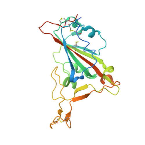
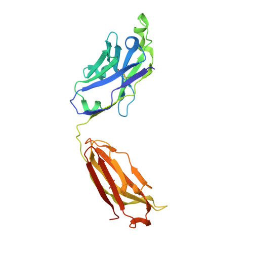
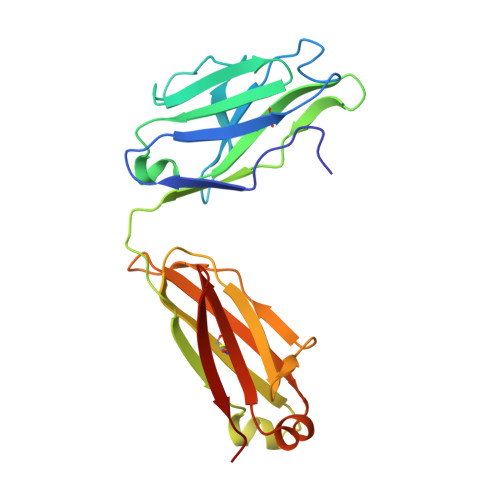
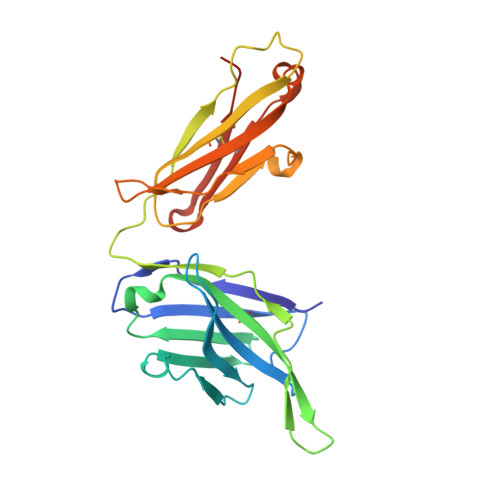
Defining a de novo non-RBM antibody as RBD-8 and its synergistic rescue of immune-evaded antibodies to neutralize Omicron SARS-CoV-2.

Rao, X.Zhao, R.Tong, Z.Guo, S.Peng, W.Liu, K.Li, S.Wu, L.Tong, J.Chai, Y.Han, P.Wang, F.Jia, P.Li, Z.Zhao, X.Li, D.Zhang, R.Zhang, X.Zou, W.Li, W.Wang, Q.Gao, G.F.Wu, Y.Dai, L.Gao, F.

(2023) Proc Natl Acad Sci U S A 120: e2314193120-e2314193120

  • DOI: https://doi.org/10.1073/pnas.2314193120
  • Primary Citation Related Structures: 
    7Y3N, 7Y3O, 8HRD

  • PubMed Abstract: 

    Currently, monoclonal antibodies (MAbs) targeting the SARS-CoV-2 receptor binding domain (RBD) of spike (S) protein are classified into seven classes based on their binding epitopes. However, most of these antibodies are seriously impaired by SARS-CoV-2 Omicron and its subvariants, especially the recent BQ.1.1, XBB and its derivatives. Identification of broadly neutralizing MAbs against currently circulating variants is imperative. In this study, we identified a "breathing" cryptic epitope in the S protein, named as RBD-8. Two human MAbs, BIOLS56 and IMCAS74, were isolated recognizing this epitope with broad neutralization abilities against tested sarbecoviruses, including SARS-CoV, pangolin-origin coronaviruses, and all the SARS-CoV-2 variants tested (Omicron BA.4/BA.5, BQ.1.1, and XBB subvariants). Searching through the literature, some more RBD-8 MAbs were defined. More importantly, BIOLS56 rescues the immune-evaded antibody, RBD-5 MAb IMCAS-L4.65, by making a bispecific MAb, to neutralize BQ.1 and BQ.1.1, thereby producing an MAb to cover all the currently circulating Omicron subvariants. Structural analysis reveals that the neutralization effect of RBD-8 antibodies depends on the extent of epitope exposure, which is affected by the angle of antibody binding and the number of up-RBDs induced by angiotensin-converting enzyme 2 binding. This cryptic epitope which recognizes non- receptor binding motif (non-RBM) provides guidance for the development of universal therapeutic antibodies and vaccines against COVID-19.


  • Organizational Affiliation
    • Laboratory of Protein Engineering and Vaccines, Tianjin Institute of Industrial Biotechnology, Chinese Academy of Sciences, Tianjin 300308, China.

Macromolecule Content 

  • Total Structure Weight: 487.97 kDa 
  • Atom Count: 33,039 
  • Modeled Residue Count: 4,316 
  • Deposited Residue Count: 4,496 
  • Unique protein chains: 5

Macromolecules

Find similar proteins by:|  3D Structure
Entity ID: 1
MoleculeChains  Sequence LengthOrganismDetailsImage
Spike protein S1A,
F [auth B],
K,
P [auth T]
223Severe acute respiratory syndrome coronavirus 2Mutation(s): 2 
Gene Names: S2
UniProt
Find proteins for P0DTC2 (Severe acute respiratory syndrome coronavirus 2)
Explore P0DTC2 
Go to UniProtKB:  P0DTC2
Entity Groups
Sequence Clusters30% Identity50% Identity70% Identity90% Identity95% Identity100% Identity
UniProt GroupP0DTC2
Glycosylation
Glycosylation Sites: 1Go to GlyGen: P0DTC2-1
Sequence Annotations
Expand
Reference Sequence
Find similar proteins by:|  3D Structure
Entity ID: 2
MoleculeChains  Sequence LengthOrganismDetailsImage
IMCAS74 Fab heavy chainB [auth O],
G [auth C],
L,
Q [auth W]
235Homo sapiensMutation(s): 0 
Find similar proteins by:|  3D Structure
Entity ID: 3
MoleculeChains  Sequence LengthOrganismDetailsImage
IMCAS74 Fab light chainC [auth P],
H [auth E],
M,
R [auth X]
217Homo sapiensMutation(s): 0 
Find similar proteins by:|  3D Structure
Entity ID: 4
MoleculeChains  Sequence LengthOrganismDetailsImage
W14 Fab heavy chainD,
I [auth G],
N,
S [auth Y]
234Homo sapiensMutation(s): 0 
Find similar proteins by:|  3D Structure
Entity ID: 5
MoleculeChains  Sequence LengthOrganismDetailsImage
W14 Fab light chainE [auth F],
J [auth H],
O [auth Q],
T [auth Z]
215Homo sapiensMutation(s): 0 

Experimental Data & Validation

Experimental Data

  • Method: X-RAY DIFFRACTION
  • Resolution: 2.86 Å
  • R-Value Free:  0.244 (Depositor), 0.245 (DCC) 
  • R-Value Work:  0.212 (Depositor), 0.213 (DCC) 
  • R-Value Observed: 0.213 (Depositor) 
Space Group: C 1 2 1
Unit Cell:
Length ( Å )Angle ( ˚ )
a = 196.662α = 90
b = 270.048β = 107.85
c = 118.156γ = 90
Software Package:
Software NamePurpose
PHENIXrefinement
PHENIXrefinement
XDSdata reduction
XDSdata scaling
PHASERphasing

Structure Validation

View Full Validation Report


Ligand Structure Quality Assessment 


Entry History 

& Funding Information

Deposition Data


Funding OrganizationLocationGrant Number
Chinese Academy of SciencesChinaXDB29010202

Revision History  (Full details and data files)

  • Version 1.0: 2023-12-20
    Type: Initial release
  • Version 1.1: 2023-12-27
    Changes: Database references
  • Version 1.2: 2024-10-23
    Changes: Structure summary