8HPK

Crystal structure of the bacterial oxalate transporter OxlT in an oxalate-bound occluded form


Experimental Data Snapshot

  • Method: X-RAY DIFFRACTION
  • Resolution: 3.00 Å
  • R-Value Free: 0.278 
  • R-Value Work: 0.237 
  • R-Value Observed: 0.241 

wwPDB Validation   3D Report Full Report


Ligand Structure Quality Assessment 


This is version 1.1 of the entry. See complete history


Literature

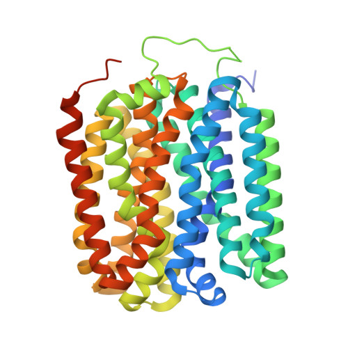
Structure and mechanism of oxalate transporter OxlT in an oxalate-degrading bacterium in the gut microbiota.

Jaunet-Lahary, T.Shimamura, T.Hayashi, M.Nomura, N.Hirasawa, K.Shimizu, T.Yamashita, M.Tsutsumi, N.Suehiro, Y.Kojima, K.Sudo, Y.Tamura, T.Iwanari, H.Hamakubo, T.Iwata, S.Okazaki, K.I.Hirai, T.Yamashita, A.

(2023) Nat Commun 14: 1730-1730

  • DOI: https://doi.org/10.1038/s41467-023-36883-5
  • Primary Citation of Related Structures:  
    8HPJ, 8HPK

  • PubMed Abstract: 

    An oxalate-degrading bacterium in the gut microbiota absorbs food-derived oxalate to use this as a carbon and energy source, thereby reducing the risk of kidney stone formation in host animals. The bacterial oxalate transporter OxlT selectively uptakes oxalate from the gut to bacterial cells with a strict discrimination from other nutrient carboxylates. Here, we present crystal structures of oxalate-bound and ligand-free OxlT in two distinct conformations, occluded and outward-facing states. The ligand-binding pocket contains basic residues that form salt bridges with oxalate while preventing the conformational switch to the occluded state without an acidic substrate. The occluded pocket can accommodate oxalate but not larger dicarboxylates, such as metabolic intermediates. The permeation pathways from the pocket are completely blocked by extensive interdomain interactions, which can be opened solely by a flip of a single side chain neighbouring the substrate. This study shows the structural basis underlying metabolic interactions enabling favourable symbiosis.


  • Organizational Affiliation

    Research Center for Computational Science, Institute for Molecular Science, National Institutes of Natural Sciences, Okazaki, 444-8585, Japan.


Macromolecules
Find similar proteins by:  (by identity cutoff)  |  3D Structure
Entity ID: 1
MoleculeChains Sequence LengthOrganismDetailsImage
Oxalate:formate antiporter427Oxalobacter formigenesMutation(s): 0 
Gene Names: oxlT
Membrane Entity: Yes 
UniProt
Find proteins for Q51330 (Oxalobacter formigenes)
Explore Q51330 
Go to UniProtKB:  Q51330
Entity Groups  
Sequence Clusters30% Identity50% Identity70% Identity90% Identity95% Identity100% Identity
UniProt GroupQ51330
Sequence Annotations
Expand
  • Reference Sequence
Find similar proteins by:  (by identity cutoff)  |  3D Structure
Entity ID: 2
MoleculeChains Sequence LengthOrganismDetailsImage
Fab fragment Heavy cheinB [auth H]220Mus musculusMutation(s): 0 
Entity Groups  
Sequence Clusters30% Identity50% Identity70% Identity90% Identity95% Identity100% Identity
Sequence Annotations
Expand
  • Reference Sequence
Find similar proteins by:  (by identity cutoff)  |  3D Structure
Entity ID: 3
MoleculeChains Sequence LengthOrganismDetailsImage
Fab fragment Light chainC [auth L]215Mus musculusMutation(s): 0 
Entity Groups  
Sequence Clusters30% Identity50% Identity70% Identity90% Identity95% Identity100% Identity
Sequence Annotations
Expand
  • Reference Sequence
Small Molecules
Ligands 1 Unique
IDChains Name / Formula / InChI Key2D Diagram3D Interactions
OXL (Subject of Investigation/LOI)
Query on OXL

Download Ideal Coordinates CCD File 
D [auth A]OXALATE ION
C2 O4
MUBZPKHOEPUJKR-UHFFFAOYSA-L
Experimental Data & Validation

Experimental Data

  • Method: X-RAY DIFFRACTION
  • Resolution: 3.00 Å
  • R-Value Free: 0.278 
  • R-Value Work: 0.237 
  • R-Value Observed: 0.241 
  • Space Group: P 21 21 2
Unit Cell:
Length ( Å )Angle ( ˚ )
a = 114.95α = 90
b = 233.19β = 90
c = 50.77γ = 90
Software Package:
Software NamePurpose
PHENIXrefinement
Cootmodel building

Structure Validation

View Full Validation Report


Ligand Structure Quality Assessment 


Entry History & Funding Information

Deposition Data


Funding OrganizationLocationGrant Number
Japan Society for the Promotion of Science (JSPS)JapanJP20H03195

Revision History  (Full details and data files)

  • Version 1.0: 2023-02-15
    Type: Initial release
  • Version 1.1: 2023-04-12
    Changes: Database references