8HG7 | pdb_00008hg7

Structure of human SGLT2-MAP17 complex with Sotagliflozin


Experimental Data Snapshot

  • Method: ELECTRON MICROSCOPY
  • Resolution: 3.10 Å
  • Aggregation State: PARTICLE 
  • Reconstruction Method: SINGLE PARTICLE 

wwPDB Validation 3D Report Full Report

Validation slider image for 8HG7

This is version 1.4 of the entry. See complete history

Literature

Transport and inhibition mechanism of the human SGLT2-MAP17 glucose transporter.

Hiraizumi, M.Akashi, T.Murasaki, K.Kishida, H.Kumanomidou, T.Torimoto, N.Nureki, O.Miyaguchi, I.

(2024) Nat Struct Mol Biol 31: 159-169

  • DOI: https://doi.org/10.1038/s41594-023-01134-0
  • Primary Citation Related Structures: 
    8HB0, 8HDH, 8HEZ, 8HG7, 8HIN

  • PubMed Abstract: 

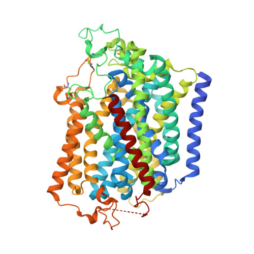
    Sodium-glucose cotransporter 2 (SGLT2) is imporant in glucose reabsorption. SGLT2 inhibitors suppress renal glucose reabsorption, therefore reducing blood glucose levels in patients with type 2 diabetes. We and others have developed several SGLT2 inhibitors starting from phlorizin, a natural product. Using cryo-electron microscopy, we present the structures of human (h)SGLT2-MAP17 complexed with five natural or synthetic inhibitors. The four synthetic inhibitors (including canagliflozin) bind the transporter in the outward conformations, while phlorizin binds it in the inward conformation. The phlorizin-hSGLT2 interaction exhibits biphasic kinetics, suggesting that phlorizin alternately binds to the extracellular and intracellular sides. The Na + -bound outward-facing and unbound inward-open structures of hSGLT2-MAP17 suggest that the MAP17-associated bundle domain functions as a scaffold, with the hash domain rotating around the Na + -binding site. Thus, Na + binding stabilizes the outward-facing conformation, and its release promotes state transition to inward-open conformation, exhibiting a role of Na + in symport mechanism. These results provide structural evidence for the Na + -coupled alternating-access mechanism proposed for the transporter family.


  • Organizational Affiliation
    • Discovery Technology Laboratories Sohyaku Innovative Research Division, Mitsubishi Tanabe Pharma, Yokohama, Japan. hiraizumi-masahiro4580@g.ecc.u-tokyo.ac.jp.

Macromolecule Content 

  • Total Structure Weight: 86.15 kDa 
  • Atom Count: 4,808 
  • Modeled Residue Count: 619 
  • Deposited Residue Count: 790 
  • Unique protein chains: 2

Macromolecules

Find similar proteins by:|  3D Structure
Entity ID: 1
MoleculeChains  Sequence LengthOrganismDetailsImage
Sodium/glucose cotransporter 2676Homo sapiensMutation(s): 0 
Gene Names: SLC5A2SGLT2
Membrane Entity: Yes 
UniProt & NIH Common Fund Data Resources
Find proteins for P31639 (Homo sapiens)
Explore P31639 
Go to UniProtKB:  P31639
PHAROS:  P31639
GTEx:  ENSG00000140675 
Entity Groups
Sequence Clusters30% Identity50% Identity70% Identity90% Identity95% Identity100% Identity
UniProt GroupP31639
Sequence Annotations
Expand
Reference Sequence
Find similar proteins by:|  3D Structure
Entity ID: 2
MoleculeChains  Sequence LengthOrganismDetailsImage
PDZK1-interacting protein 1114Homo sapiensMutation(s): 0 
Gene Names: MAP17
Membrane Entity: Yes 
UniProt & NIH Common Fund Data Resources
Find proteins for Q13113 (Homo sapiens)
Explore Q13113 
Go to UniProtKB:  Q13113
PHAROS:  Q13113
GTEx:  ENSG00000162366 
Entity Groups
Sequence Clusters30% Identity50% Identity70% Identity90% Identity95% Identity100% Identity
UniProt GroupQ13113
Sequence Annotations
Expand
Reference Sequence

Small Molecules

Ligands 3 Unique
IDChains Name / Formula / InChI Key2D Diagram3D Interactions
LFL
(Subject of Investigation/LOI)

Query on LFL


Download:Ideal Coordinates CCD File
D [auth A](2S,3R,4R,5S,6R)-2-[4-chloranyl-3-[(4-ethoxyphenyl)methyl]phenyl]-6-methylsulfanyl-oxane-3,4,5-triol
C21 H25 Cl O5 S
QKDRXGFQVGOQKS-CRSSMBPESA-N
NAG

Query on NAG


Download:Ideal Coordinates CCD File
C [auth A]2-acetamido-2-deoxy-beta-D-glucopyranose
C8 H15 N O6
OVRNDRQMDRJTHS-FMDGEEDCSA-N
NA

Query on NA


Download:Ideal Coordinates CCD File
E [auth A]SODIUM ION
Na
FKNQFGJONOIPTF-UHFFFAOYSA-N
Binding Affinity Annotations 
IDSourceBinding Affinity
LFL BindingDB:  8HG7 IC50: min: 1.5, max: 1.8 (nM) from 2 assay(s)

Experimental Data & Validation

Experimental Data

  • Method: ELECTRON MICROSCOPY
  • Resolution: 3.10 Å
  • Aggregation State: PARTICLE 
  • Reconstruction Method: SINGLE PARTICLE 

Structure Validation

View Full Validation Report


Entry History 

& Funding Information

Deposition Data


Funding OrganizationLocationGrant Number
Other private--

Revision History  (Full details and data files)

  • Version 1.0: 2023-11-15
    Type: Initial release
  • Version 1.1: 2023-12-13
    Changes: Database references
  • Version 1.2: 2023-12-20
    Changes: Database references
  • Version 1.3: 2024-02-07
    Changes: Database references
  • Version 1.4: 2024-10-16
    Changes: Data collection, Structure summary