8HBL | pdb_00008hbl

Crystal structure of the SARS-unique domain (SUD) of SARS-CoV-2 (1.58 angstrom resolution)


Experimental Data Snapshot

  • Method: X-RAY DIFFRACTION
  • Resolution: 1.58 Å
  • R-Value Free: 
    0.180 (Depositor), 0.181 (DCC) 
  • R-Value Work: 
    0.139 (Depositor), 0.141 (DCC) 
  • R-Value Observed: 
    0.141 (Depositor) 

Starting Model: experimental
View more details

wwPDB Validation 3D Report Full Report

Validation slider image for 8HBL

Ligand Structure Quality Assessment 


This is version 1.3 of the entry. See complete history

Literature

Identification of the SARS-unique domain of SARS-CoV-2 as an antiviral target.

Qin, B.Li, Z.Tang, K.Wang, T.Xie, Y.Aumonier, S.Wang, M.Yuan, S.Cui, S.

(2023) Nat Commun 14: 3999-3999

  • DOI: https://doi.org/10.1038/s41467-023-39709-6
  • Primary Citation Related Structures: 
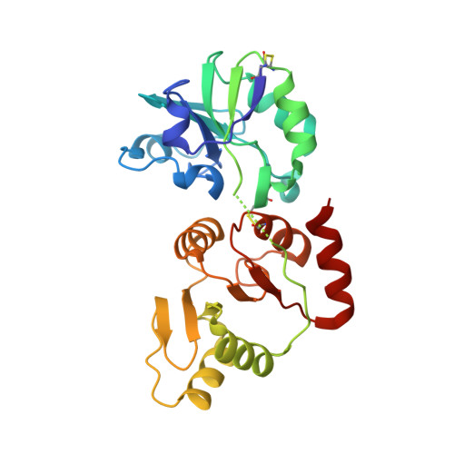
    8GQC, 8HBL

  • PubMed Abstract: 

    SARS-CoV-2 nsp3 is essential for viral replication and host responses. The SARS-unique domain (SUD) of nsp3 exerts its function through binding to viral and host proteins and RNAs. Herein, we show that SARS-CoV-2 SUD is highly flexible in solution. The intramolecular disulfide bond of SARS-CoV SUD is absent in SARS-CoV-2 SUD. Incorporating this bond in SARS-CoV-2 SUD allowed crystal structure determination to 1.35 Å resolution. However, introducing this bond in SARS-CoV-2 genome was lethal for the virus. Using biolayer interferometry, we screened compounds directly binding to SARS-CoV-2 SUD and identified theaflavin 3,3'-digallate (TF3) as a potent binder, K d 2.8 µM. TF3 disrupted the SUD-guanine quadruplex interactions and exhibited anti-SARS-CoV-2 activity in Vero E6-TMPRSS2 cells with an EC 50 of 5.9 µM and CC 50 of 98.5 µM. In this work, we provide evidence that SARS-CoV-2 SUD harbors druggable sites for antiviral development.


  • Organizational Affiliation
    • NHC Key Laboratory of Systems Biology of Pathogens, Institute of Pathogen Biology Chinese Academy of Medical Sciences & Peking Union Medical College, 100730, Beijing, China.

Macromolecule Content 

  • Total Structure Weight: 29.05 kDa 
  • Atom Count: 2,201 
  • Modeled Residue Count: 252 
  • Deposited Residue Count: 260 
  • Unique protein chains: 1

Macromolecules

Find similar proteins by:|  3D Structure
Entity ID: 1
MoleculeChains  Sequence LengthOrganismDetailsImage
Non-structural protein 3260Severe acute respiratory syndrome coronavirus 2Mutation(s): 2 
UniProt
Find proteins for P0DTD1 (Severe acute respiratory syndrome coronavirus 2)
Explore P0DTD1 
Go to UniProtKB:  P0DTD1
Entity Groups
Sequence Clusters30% Identity50% Identity70% Identity90% Identity95% Identity100% Identity
UniProt GroupP0DTD1
Sequence Annotations
Expand
Reference Sequence

Small Molecules

Experimental Data & Validation

Experimental Data

  • Method: X-RAY DIFFRACTION
  • Resolution: 1.58 Å
  • R-Value Free:  0.180 (Depositor), 0.181 (DCC) 
  • R-Value Work:  0.139 (Depositor), 0.141 (DCC) 
  • R-Value Observed: 0.141 (Depositor) 
Space Group: P 31 2 1
Unit Cell:
Length ( Å )Angle ( ˚ )
a = 85.671α = 90
b = 85.671β = 90
c = 76.662γ = 120
Software Package:
Software NamePurpose
XSCALEdata scaling
PHENIXrefinement
PDB_EXTRACTdata extraction
XDSdata reduction
PHENIXphasing

Structure Validation

View Full Validation Report


Ligand Structure Quality Assessment 


Entry History 

& Funding Information

Deposition Data


Funding OrganizationLocationGrant Number
National Natural Science Foundation of China (NSFC)China81772107

Revision History  (Full details and data files)

  • Version 1.0: 2023-07-12
    Type: Initial release
  • Version 1.1: 2023-11-29
    Changes: Data collection, Refinement description
  • Version 1.2: 2024-02-14
    Changes: Database references
  • Version 1.3: 2024-10-30
    Changes: Structure summary