


Cataract-causing Y204X mutation of crystallin protein CRY beta B1 promotes its C-terminal degradation and higher-order oligomerization.
Jing, X., Zhu, M., Lu, X., Wei, P., Shi, L., Zhang, B.Y., Xu, Y., Tang, Y.P., Xiang, D.M., Gong, P.(2023) J Biological Chem 299: 104953-104953
- PubMed: 37356717 Search on PubMedSearch on PubMed Central
- DOI: https://doi.org/10.1016/j.jbc.2023.104953
- Primary Citation Related Structures:
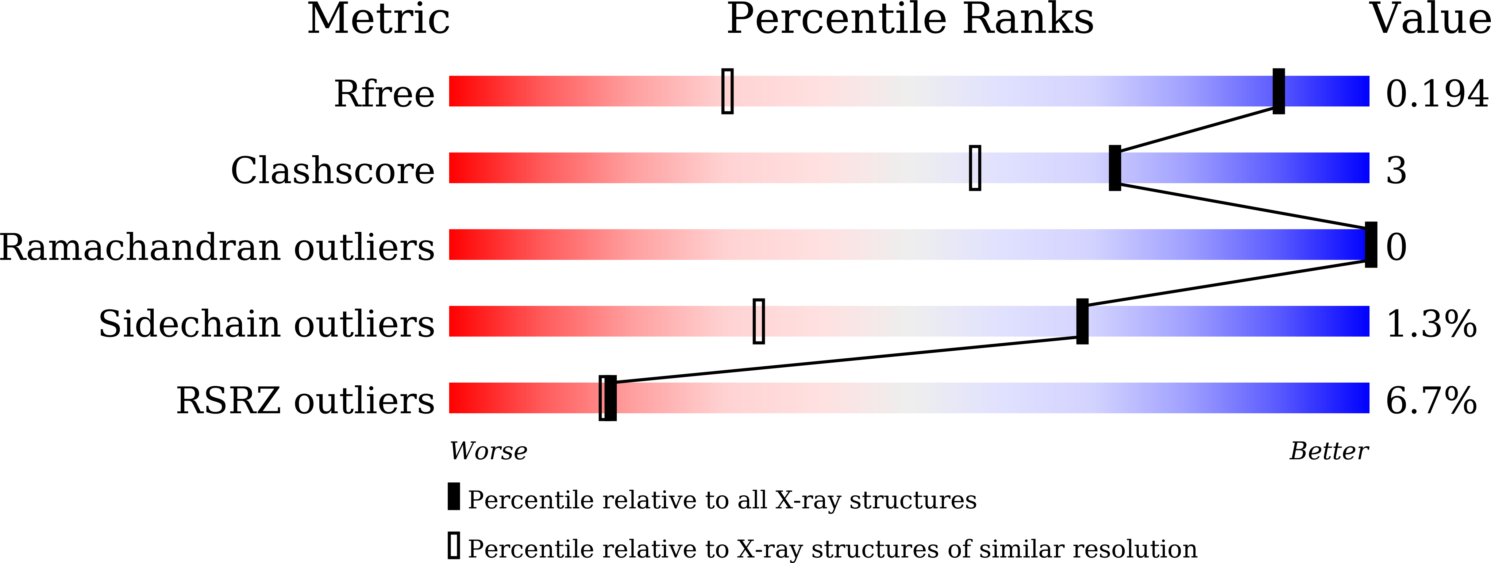
8H0R - PubMed Abstract:
Crystallin proteins are a class of main structural proteins of the vertebrate eye lens, and their solubility and stability directly determine transparency and refractive power of the lens. Mutation in genes that encode these crystallin proteins is the most common cause for congenital cataracts. Despite extensive studies, the pathogenic and molecular mechanisms that effect congenital cataracts remain unclear. In this study, we identified a novel mutation in CRYBB1 from a congenital cataract family, and demonstrated that this mutation led to an early termination of mRNA translation, resulting in a 49-residue C-terminally truncated CRYβB1 protein. We show this mutant is susceptible to proteolysis, which allowed us to determine a 1.2-Å resolution crystal structure of CRYβB1 without the entire C-terminal domain. In this crystal lattice, we observed that two N-terminal domain monomers form a dimer that structurally resembles the WT monomer, but with different surface characteristics. Biochemical analyses and cell-based data also suggested that this mutant is significantly more liable to aggregate and degrade compared to WT CRYβB1. Taken together, our results provide an insight into the mechanism regarding how a mutant crystalin contributes to the development of congenital cataract possibly through alteration of inter-protein interactions that result in protein aggregation.
- Joint Laboratory for Translational Precision Medicine, Guangzhou Women and Children's Medical Center, Guangzhou Medical University, Guangzhou, Guangdong, China; Joint Laboratory for Translational Precision Medicine, Wuhan Institute of Virology, Chinese Academy of Sciences, Wuhan, Hubei, China; Key Laboratory of Special Pathogens and Biosafety, Wuhan Institute of Virology, Center for Biosafety Mega-Science, Chinese Academy of Sciences, Wuhan, Hubei, China.
Organizational Affiliation:


