8GYR

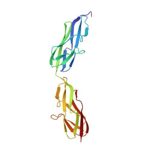
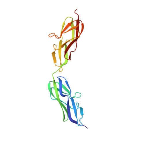
Crystal structure of a variable region segment of Leptospira host-interacting outer surface protein, LigA


Experimental Data Snapshot

  • Method: X-RAY DIFFRACTION
  • Resolution: 1.87 Å
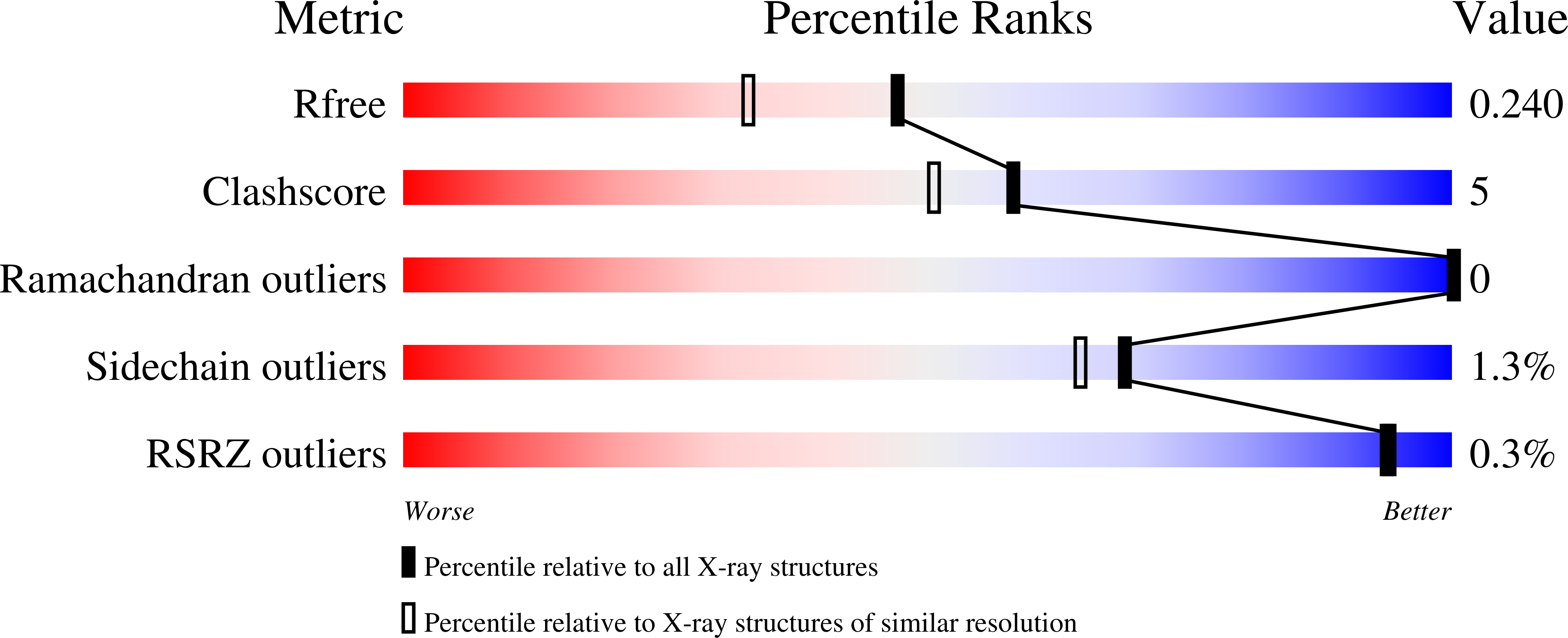
  • R-Value Free: 0.234 
  • R-Value Work: 0.208 
  • R-Value Observed: 0.209 

Starting Model: experimental
View more details

wwPDB Validation   3D Report Full Report


This is version 1.2 of the entry. See complete history


Literature

Crystal structure of a variable region segment of Leptospira host-interacting outer surface protein, LigA, reveals the orientation of Ig-like domains.

Kumar, P.Vyas, P.Faisal, S.M.Chang, Y.F.Akif, M.

(2023) Int J Biol Macromol 244: 125445-125445

  • DOI: https://doi.org/10.1016/j.ijbiomac.2023.125445
  • Primary Citation of Related Structures:  
    8GYR

  • PubMed Abstract: 

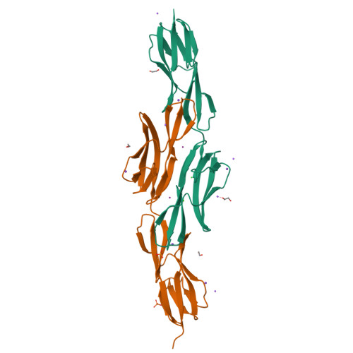
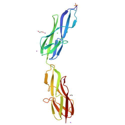
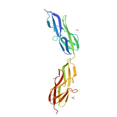
    Leptospiral immunoglobulin-like (Lig) protein family is a surface-exposed protein from the pathogenic Leptospira. The Lig protein family has been identified as an essential virulence factor of L. interrogan. One of the family members, LigA, contains 13 homologous tandem repeats of bacterial Ig-like (Big) domains in its extracellular portion. It is crucial in binding with the host's Extracellular matrices (ECM) and complement factors. However, its vital role in the invasion and evasion of pathogenic Leptospira, structural details, and domain organization of the extracellular portion of this protein are not explored thoroughly. Here, we described the first high-resolution crystal structure of a variable region segment (LigA8-9) of LigA at 1.87 Å resolution. The structure showed some remarkably distinctive aspects compared with other closely related Immunoglobulin domains. The structure illustrated the relative orientation of two domains and highlighted the role of the linker region in the domain orientation. We also observed an apparent electron density of Ca 2+ ions coordinated with a proper interacting geometry within the protein. Molecular dynamic simulations demonstrated the involvement of a linker salt bridge in providing rigidity between the two domains. Our study proposes an overall arrangement of Ig-like domains in the LigA protein. The structural understanding of the extracellular portion of LigA and its interaction with the ECM provides insight into developing new therapeutics directed toward leptospirosis.


  • Organizational Affiliation

    Laboratory of Structural Biology, Department of Biochemistry, School of Life Sciences, University of Hyderabad, Gachibowli, Hyderabad, India.


Macromolecules
Find similar proteins by:  (by identity cutoff)  |  3D Structure
Entity ID: 1
MoleculeChains Sequence LengthOrganismDetailsImage
Leptospira Immunoglobulin-like protein182Leptospira interrogansMutation(s): 0 
UniProt
Find proteins for C0J1Q2 (Leptospira interrogans)
Explore C0J1Q2 
Go to UniProtKB:  C0J1Q2
Entity Groups  
Sequence Clusters30% Identity50% Identity70% Identity90% Identity95% Identity100% Identity
UniProt GroupC0J1Q2
Sequence Annotations
Expand
  • Reference Sequence
Find similar proteins by:  (by identity cutoff)  |  3D Structure
Entity ID: 2
MoleculeChains Sequence LengthOrganismDetailsImage
LigA183Leptospira interrogansMutation(s): 0 
Gene Names: ligA
UniProt
Find proteins for C0J1Q2 (Leptospira interrogans)
Explore C0J1Q2 
Go to UniProtKB:  C0J1Q2
Entity Groups  
Sequence Clusters30% Identity50% Identity70% Identity90% Identity95% Identity100% Identity
UniProt GroupC0J1Q2
Sequence Annotations
Expand
  • Reference Sequence
Small Molecules
Ligands 7 Unique
IDChains Name / Formula / InChI Key2D Diagram3D Interactions
IOD
Query on IOD

Download Ideal Coordinates CCD File 
L [auth A],
O [auth B]
IODIDE ION
I
XMBWDFGMSWQBCA-UHFFFAOYSA-M
PO4
Query on PO4

Download Ideal Coordinates CCD File 
H [auth A],
T [auth B]
PHOSPHATE ION
O4 P
NBIIXXVUZAFLBC-UHFFFAOYSA-K
EDO
Query on EDO

Download Ideal Coordinates CCD File 
F [auth A],
G [auth A],
R [auth B],
S [auth B]
1,2-ETHANEDIOL
C2 H6 O2
LYCAIKOWRPUZTN-UHFFFAOYSA-N
CA
Query on CA

Download Ideal Coordinates CCD File 
K [auth A],
N [auth B]
CALCIUM ION
Ca
BHPQYMZQTOCNFJ-UHFFFAOYSA-N
K
Query on K

Download Ideal Coordinates CCD File 
C [auth A],
D [auth A],
P [auth B],
Q [auth B],
V [auth B]
POTASSIUM ION
K
NPYPAHLBTDXSSS-UHFFFAOYSA-N
CL
Query on CL

Download Ideal Coordinates CCD File 
E [auth A]CHLORIDE ION
Cl
VEXZGXHMUGYJMC-UHFFFAOYSA-M
NA
Query on NA

Download Ideal Coordinates CCD File 
I [auth A],
J [auth A],
M [auth A],
U [auth B],
W [auth B]
SODIUM ION
Na
FKNQFGJONOIPTF-UHFFFAOYSA-N
Experimental Data & Validation

Experimental Data

  • Method: X-RAY DIFFRACTION
  • Resolution: 1.87 Å
  • R-Value Free: 0.234 
  • R-Value Work: 0.208 
  • R-Value Observed: 0.209 
  • Space Group: P 21 21 21
Unit Cell:
Length ( Å )Angle ( ˚ )
a = 34.219α = 90
b = 63.905β = 90
c = 171.839γ = 90
Software Package:
Software NamePurpose
REFMACrefinement
DIALSdata reduction
pointlessdata scaling
BUCCANEERphasing

Structure Validation

View Full Validation Report


Entry History & Funding Information

Deposition Data


Funding OrganizationLocationGrant Number
Science and Engineering Research Board (SERB)IndiaEMR/2016/001183

Revision History  (Full details and data files)

  • Version 1.0: 2023-06-28
    Type: Initial release
  • Version 1.1: 2023-07-05
    Changes: Database references
  • Version 1.2: 2024-02-07
    Changes: Data collection, Refinement description