Primary Citation Related Structures: 7XGY, 7YIM, 8GVK
PubMed Abstract:
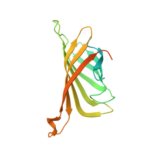
Cryo-electron microscopy (cryo-EM) visualizes the atomic structure of macromolecules that are embedded in vitrified thin ice at their close-to-native state. However, the homogeneity of ice thickness, a key factor to ensure high image quality, is poorly controlled during specimen preparation and has become one of the main challenges for high-resolution cryo-EM. Here we found that the uniformity of thin ice relies on the surface flatness of the supporting film, and developed a method to use ultraflat graphene (UFG) as the support for cryo-EM specimen preparation to achieve better control of vitreous ice thickness. We show that the uniform thin ice on UFG improves the image quality of vitrified specimens. Using such a method we successfully determined the three-dimensional structures of hemoglobin (64 kDa), α-fetoprotein (67 kDa) with no symmetry, and streptavidin (52 kDa) at a resolution of 3.5 Å, 2.6 Å and 2.2 Å, respectively. Furthermore, our results demonstrate the potential of UFG for the fields of cryo-electron tomography and structure-based drug discovery.
Organizational Affiliation:
Center for Nanochemistry, Beijing Science and Engineering Center for Nanocarbons, Beijing National Laboratory for Molecular Sciences, College of Chemistry and Molecular Engineering, Peking University, Beijing, China.
Beijing Graphene Institute (BGI), Beijing, China.
Ministry of Education Key Laboratory of Protein Sciences, Beijing Frontier Research Center for Biological Structures, Beijing Advanced Innovation Center for Structural Biology, School of Life Sciences, Tsinghua University, Beijing, China. nanliuem@tsinghua.edu.cn.
State Key Laboratory for Turbulence and Complex System, Department of Mechanics and Engineering Science, College of Engineering, Peking University, Beijing, China.
Hainan Provincial Key Laboratory of Carcinogenesis and Intervention, Hainan Medical College, Haikou, China.
Department of Biology, School of Life Sciences, Southern University of Science and Technology, Shenzhen, China.
Academy for Advanced Interdisciplinary Studies, Peking University, Beijing, China.
Ministry of Education Key Laboratory of Protein Sciences, Beijing Frontier Research Center for Biological Structures, Beijing Advanced Innovation Center for Structural Biology, School of Life Sciences, Tsinghua University, Beijing, China.
Tsinghua-Peking Joint Center for Life Sciences, Tsinghua University, Beijing, China.
State Key Laboratory for Turbulence and Complex System, Department of Mechanics and Engineering Science, College of Engineering, Peking University, Beijing, China. xdwei@pku.edu.cn.
Beijing Innovation Center for Engineering Science and Advanced Technology, Peking University, Beijing, China. xdwei@pku.edu.cn.
Peking University Nanchang Innovation Institute, Nanchang, China. xdwei@pku.edu.cn.
Ministry of Education Key Laboratory of Protein Sciences, Beijing Frontier Research Center for Biological Structures, Beijing Advanced Innovation Center for Structural Biology, School of Life Sciences, Tsinghua University, Beijing, China. hongweiwang@tsinghua.edu.cn.
Tsinghua-Peking Joint Center for Life Sciences, Tsinghua University, Beijing, China. hongweiwang@tsinghua.edu.cn.
Center for Nanochemistry, Beijing Science and Engineering Center for Nanocarbons, Beijing National Laboratory for Molecular Sciences, College of Chemistry and Molecular Engineering, Peking University, Beijing, China. hlpeng@pku.edu.cn.
Beijing Graphene Institute (BGI), Beijing, China. hlpeng@pku.edu.cn.
Academy for Advanced Interdisciplinary Studies, Peking University, Beijing, China. hlpeng@pku.edu.cn.