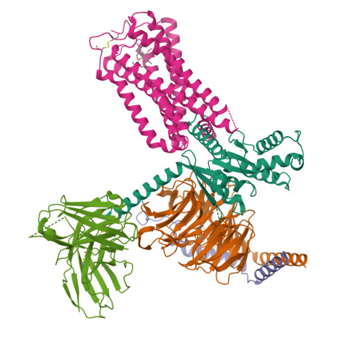
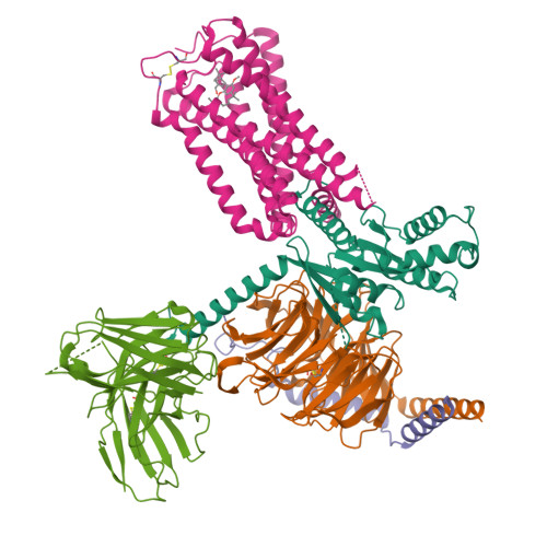
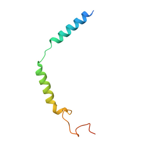
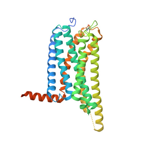
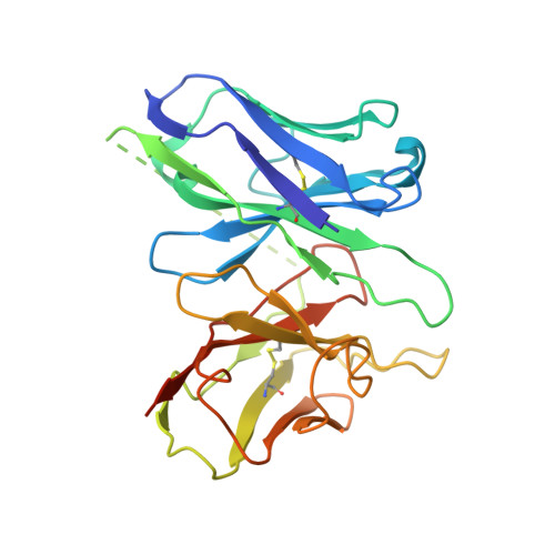
8GUS

Cryo-EM structure of HU-CB2-G protein complex


Experimental Data Snapshot

  • Method: ELECTRON MICROSCOPY
  • Resolution: 2.97 Å
  • Aggregation State: PARTICLE 
  • Reconstruction Method: SINGLE PARTICLE 

Starting Model: experimental
View more details

wwPDB Validation   3D Report Full Report


This is version 1.0 of the entry. See complete history


Literature

Structural basis of selective cannabinoid CB 2 receptor activation.

Li, X.Chang, H.Bouma, J.de Paus, L.V.Mukhopadhyay, P.Paloczi, J.Mustafa, M.van der Horst, C.Kumar, S.S.Wu, L.Yu, Y.van den Berg, R.J.B.H.N.Janssen, A.P.A.Lichtman, A.Liu, Z.J.Pacher, P.van der Stelt, M.Heitman, L.H.Hua, T.

(2023) Nat Commun 14: 1447-1447

  • DOI: https://doi.org/10.1038/s41467-023-37112-9
  • Primary Citation of Related Structures:  
    8GUQ, 8GUR, 8GUS, 8GUT

  • PubMed Abstract: 

    Cannabinoid CB 2 receptor (CB 2 R) agonists are investigated as therapeutic agents in the clinic. However, their molecular mode-of-action is not fully understood. Here, we report the discovery of LEI-102, a CB 2 R agonist, used in conjunction with three other CBR ligands (APD371, HU308, and CP55,940) to investigate the selective CB 2 R activation by binding kinetics, site-directed mutagenesis, and cryo-EM studies. We identify key residues for CB 2 R activation. Highly lipophilic HU308 and the endocannabinoids, but not the more polar LEI-102, APD371, and CP55,940, reach the binding pocket through a membrane channel in TM1-TM7. Favorable physico-chemical properties of LEI-102 enable oral efficacy in a chemotherapy-induced nephropathy model. This study delineates the molecular mechanism of CB 2 R activation by selective agonists and highlights the role of lipophilicity in CB 2 R engagement. This may have implications for GPCR drug design and sheds light on their activation by endogenous ligands.


  • Organizational Affiliation

    iHuman Institute, ShanghaiTech University, Shanghai, 201210, China.


Macromolecules
Find similar proteins by:  (by identity cutoff)  |  3D Structure
Entity ID: 1
MoleculeChains Sequence LengthOrganismDetailsImage
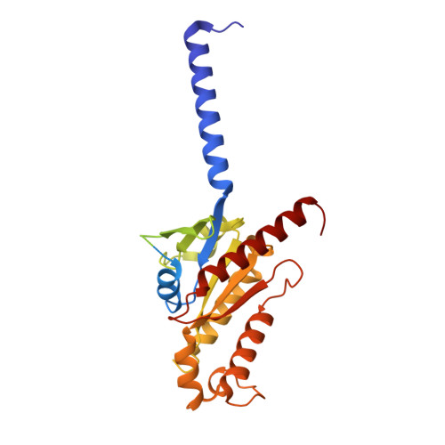
Guanine nucleotide-binding protein G(i) subunit alpha-1354Homo sapiensMutation(s): 0 
Gene Names: GNAI1
EC: 3.6.5
Membrane Entity: Yes 
UniProt & NIH Common Fund Data Resources
Find proteins for P63096 (Homo sapiens)
Explore P63096 
Go to UniProtKB:  P63096
PHAROS:  P63096
GTEx:  ENSG00000127955 
Entity Groups  
Sequence Clusters30% Identity50% Identity70% Identity90% Identity95% Identity100% Identity
UniProt GroupP63096
Sequence Annotations
Expand
  • Reference Sequence
Find similar proteins by:  (by identity cutoff)  |  3D Structure
Entity ID: 2
MoleculeChains Sequence LengthOrganismDetailsImage
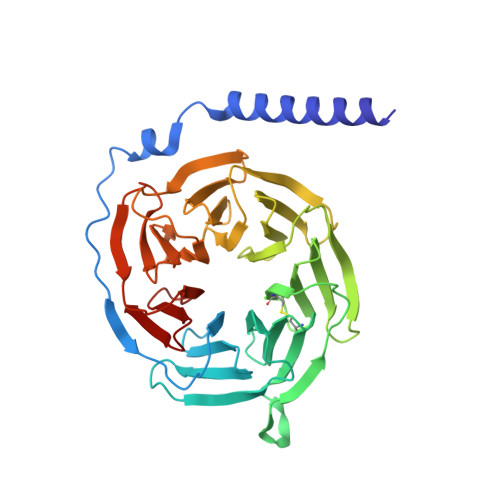
Guanine nucleotide-binding protein G(I)/G(S)/G(T) subunit beta-1340Homo sapiensMutation(s): 0 
Gene Names: GNB1
UniProt & NIH Common Fund Data Resources
Find proteins for P62873 (Homo sapiens)
Explore P62873 
Go to UniProtKB:  P62873
PHAROS:  P62873
GTEx:  ENSG00000078369 
Entity Groups  
Sequence Clusters30% Identity50% Identity70% Identity90% Identity95% Identity100% Identity
UniProt GroupP62873
Sequence Annotations
Expand
  • Reference Sequence
Find similar proteins by:  (by identity cutoff)  |  3D Structure
Entity ID: 3
MoleculeChains Sequence LengthOrganismDetailsImage
Guanine nucleotide-binding protein G(I)/G(S)/G(O) subunit gamma-271Homo sapiensMutation(s): 0 
Gene Names: GNG2
Membrane Entity: Yes 
UniProt & NIH Common Fund Data Resources
Find proteins for P59768 (Homo sapiens)
Explore P59768 
Go to UniProtKB:  P59768
PHAROS:  P59768
GTEx:  ENSG00000186469 
Entity Groups  
Sequence Clusters30% Identity50% Identity70% Identity90% Identity95% Identity100% Identity
UniProt GroupP59768
Sequence Annotations
Expand
  • Reference Sequence
Find similar proteins by:  (by identity cutoff)  |  3D Structure
Entity ID: 4
MoleculeChains Sequence LengthOrganismDetailsImage
Cannabinoid receptor 2D [auth R]360Homo sapiensMutation(s): 0 
Gene Names: CNR2CB2ACB2B
Membrane Entity: Yes 
UniProt & NIH Common Fund Data Resources
Find proteins for P34972 (Homo sapiens)
Explore P34972 
Go to UniProtKB:  P34972
PHAROS:  P34972
GTEx:  ENSG00000188822 
Entity Groups  
Sequence Clusters30% Identity50% Identity70% Identity90% Identity95% Identity100% Identity
UniProt GroupP34972
Sequence Annotations
Expand
  • Reference Sequence
Find similar proteins by:  (by identity cutoff)  |  3D Structure
Entity ID: 5
MoleculeChains Sequence LengthOrganismDetailsImage
scFv16E [auth S]259Homo sapiensMutation(s): 0 
Entity Groups  
Sequence Clusters30% Identity50% Identity70% Identity90% Identity95% Identity100% Identity
Sequence Annotations
Expand
  • Reference Sequence
Small Molecules
Ligands 1 Unique
IDChains Name / Formula / InChI Key2D Diagram3D Interactions
KO3 (Subject of Investigation/LOI)
Query on KO3

Download Ideal Coordinates CCD File 
F [auth R][(1~{S},4~{S},5~{S})-4-[2,6-dimethoxy-4-(2-methyloctan-2-yl)phenyl]-6,6-dimethyl-2-bicyclo[3.1.1]hept-2-enyl]methanol
C27 H42 O3
CFMRIVODIXTERW-BDTNDASRSA-N
Binding Affinity Annotations 
IDSourceBinding Affinity
KO3 BindingDB:  8GUS Ki: min: 11, max: 44 (nM) from 3 assay(s)
EC50: min: 76, max: 150 (nM) from 5 assay(s)
Experimental Data & Validation

Experimental Data

  • Method: ELECTRON MICROSCOPY
  • Resolution: 2.97 Å
  • Aggregation State: PARTICLE 
  • Reconstruction Method: SINGLE PARTICLE 
EM Software:
TaskSoftware PackageVersion
MODEL REFINEMENTPHENIX

Structure Validation

View Full Validation Report


Entry History & Funding Information

Deposition Data


Funding OrganizationLocationGrant Number
National Science Foundation (NSF, China)China32022038
National Natural Science Foundation of China (NSFC)China31870744

Revision History  (Full details and data files)

  • Version 1.0: 2023-05-10
    Type: Initial release