8GTR

CryoEM structure of human Pannexin isoform 3


Experimental Data Snapshot

  • Method: ELECTRON MICROSCOPY
  • Resolution: 3.91 Å
  • Aggregation State: PARTICLE 
  • Reconstruction Method: SINGLE PARTICLE 

wwPDB Validation   3D Report Full Report


This is version 1.1 of the entry. See complete history


Literature

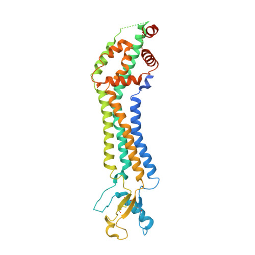
Cryo-EM structures of pannexin 1 and 3 reveal differences among pannexin isoforms.

Hussain, N.Apotikar, A.Pidathala, S.Mukherjee, S.Burada, A.P.Sikdar, S.K.Vinothkumar, K.R.Penmatsa, A.

(2024) Nat Commun 15: 2942-2942

  • DOI: https://doi.org/10.1038/s41467-024-47142-6
  • Primary Citation of Related Structures:  
    8GTR, 8GTS, 8GTT

  • PubMed Abstract: 

    Pannexins are single-membrane large-pore channels that release ions and ATP upon activation. Three isoforms of pannexins 1, 2, and 3, perform diverse cellular roles and differ in their pore lining residues. In this study, we report the cryo-EM structure of pannexin 3 at 3.9 Å and analyze its structural differences with pannexin isoforms 1 and 2. The pannexin 3 vestibule has two distinct chambers and a wider pore radius in comparison to pannexins 1 and 2. We further report two cryo-EM structures of pannexin 1, with pore substitutions W74R/R75D that mimic the pore lining residues of pannexin 2 and a germline mutant of pannexin 1, R217H at resolutions of 3.2 Å and 3.9 Å, respectively. Substitution of cationic residues in the vestibule of pannexin 1 results in reduced ATP interaction propensities to the channel. The germline mutant R217H in transmembrane helix 3 (TM3), leads to a partially constricted pore, reduced ATP interaction and weakened voltage sensitivity. The study compares the three pannexin isoform structures, the effects of substitutions of pore and vestibule-lining residues and allosteric effects of a pathological substitution on channel structure and function thereby enhancing our understanding of this vital group of ATP-release channels.


  • Organizational Affiliation

    Molecular Biophysics Unit, Indian Institute of Science, Bangalore, 560012, India.


Macromolecules
Find similar proteins by:  (by identity cutoff)  |  3D Structure
Entity ID: 1
MoleculeChains Sequence LengthOrganismDetailsImage
Pannexin-3
A, B, C, D, E
A, B, C, D, E, F, G
392Homo sapiensMutation(s): 0 
Gene Names: PANX3
Membrane Entity: Yes 
UniProt & NIH Common Fund Data Resources
Find proteins for Q96QZ0 (Homo sapiens)
Explore Q96QZ0 
Go to UniProtKB:  Q96QZ0
PHAROS:  Q96QZ0
GTEx:  ENSG00000154143 
Entity Groups  
Sequence Clusters30% Identity50% Identity70% Identity90% Identity95% Identity100% Identity
UniProt GroupQ96QZ0
Sequence Annotations
Expand
  • Reference Sequence
Small Molecules
Ligands 2 Unique
IDChains Name / Formula / InChI Key2D Diagram3D Interactions
PTY
Query on PTY

Download Ideal Coordinates CCD File 
I [auth A]
K [auth B]
M [auth C]
N [auth D]
P [auth E]
I [auth A],
K [auth B],
M [auth C],
N [auth D],
P [auth E],
R [auth F],
T [auth G]
PHOSPHATIDYLETHANOLAMINE
C40 H80 N O8 P
NJGIRBISCGPRPF-KXQOOQHDSA-N
NAG
Query on NAG

Download Ideal Coordinates CCD File 
H [auth A]
J [auth B]
L [auth C]
O [auth D]
Q [auth E]
H [auth A],
J [auth B],
L [auth C],
O [auth D],
Q [auth E],
S [auth F],
U [auth G]
2-acetamido-2-deoxy-beta-D-glucopyranose
C8 H15 N O6
OVRNDRQMDRJTHS-FMDGEEDCSA-N
Experimental Data & Validation

Experimental Data

  • Method: ELECTRON MICROSCOPY
  • Resolution: 3.91 Å
  • Aggregation State: PARTICLE 
  • Reconstruction Method: SINGLE PARTICLE 
EM Software:
TaskSoftware PackageVersion
RECONSTRUCTIONcryoSPARC
MODEL REFINEMENTPHENIX

Structure Validation

View Full Validation Report


Entry History & Funding Information

Deposition Data


Funding OrganizationLocationGrant Number
Department of Biotechnology (DBT, India)IndiaIA/I/15/2/502063

Revision History  (Full details and data files)

  • Version 1.0: 2024-02-28
    Type: Initial release
  • Version 1.1: 2024-06-19
    Changes: Database references