Improvement of substrate specificity of the direct electron transfer type FAD-dependent glucose dehydrogenase catalytic subunit.
Kerrigan Jr., J.A., Yoshida, H., Okuda-Shimazaki, J., Temple, B., Kojima, K., Sode, K.(2024) J Biotechnol
- PubMed: 39326560 Search on PubMed
- DOI: https://doi.org/10.1016/j.jbiotec.2024.09.013
- Primary Citation Related Structures:
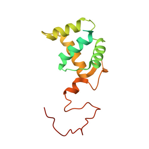
8GRJ - PubMed Abstract:
The heterotrimeric flavin adenine dinucleotide (FAD) dependent glucose dehydrogenase derived from Burkholderia cepacia (BcGDH) has many exceptional features for its use in glucose sensing-including that this enzyme is capable of direct electron transfer with an electrode in its heterotrimeric configuration. However, this enzyme's high catalytic activity towards not only glucose but also galactose presents an engineering challenge. To increase the substrate specificity of this enzyme, it must be engineered to reduce its activity towards galactose while maintaining its activity towards glucose. To aid in these mutagenesis studies, the crystal structure composed of BcGDH's small subunit and catalytic subunit (BcGDHγα), in complex with D-glucono-1,5-lactone was elucidated and used to construct the three-dimensional model for targeted, site-directed mutagenesis. BcGDHγα was then mutated at three different residues, glycine 322, asparagine 474 and asparagine 475. The single mutations that showed the greatest glucose selectivity were combined to create the resulting mutant, α-G322Q-N474S-N475S. The α-G322Q-N474S-N475S mutant and BcGDHγα wild type were then characterized with dye-mediated dehydrogenase activity assays to determine their kinetic parameters. The α-G322Q-N474S-N475S mutant showed more than a 2-fold increase in V max towards glucose and this mutant showed a lower activity towards galactose in the physiological range (5 mM) of 4.19 U mg -1 , as compared to the wild type, 86.6 U mg -1 . This resulting increase in specificity lead to an 81.7 gal/glc % activity for the wild type while the α-G322Q-N474S-N475S mutant had just 10.9 gal/glc % activity at 5 mM. While the BcGDHγα wild type has high specificity towards galactose, our engineering α-G322Q-N474S-N475S mutant showed concentration dependent response to glucose and was not affected by galactose.
- Joint Department of Biomedical Engineering, The University of North Carolina at Chapel Hill and North Carolina State University, Chapel Hill, NC 27599, USA.
Organizational Affiliation: