8GME

Crystal structure of the gp32-Dda-dT17 complex


Experimental Data Snapshot

  • Method: X-RAY DIFFRACTION
  • Resolution: 4.98 Å
  • R-Value Free: 
    0.313 (Depositor), 0.340 (DCC) 
  • R-Value Work: 
    0.286 (Depositor), 0.320 (DCC) 
  • R-Value Observed: 
    0.297 (Depositor) 

Starting Models: experimental
View more details

wwPDB Validation   3D Report Full Report


This is version 3.3 of the entry. See complete history


Literature

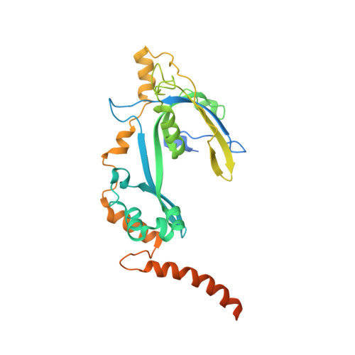
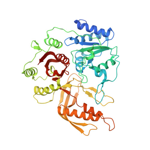
Structural and functional insights into the interaction between the bacteriophage T4 DNA processing proteins gp32 and Dda.

He, X.Yun, M.K.Li, Z.Waddell, M.B.Nourse, A.Churion, K.A.Kreuzer, K.N.Byrd, A.K.White, S.W.

(2024) Nucleic Acids Res 52: 12748-12762

  • DOI: https://doi.org/10.1093/nar/gkae910
  • Primary Citation of Related Structures:  
    8GME, 8S9I

  • PubMed Abstract: 

    Bacteriophage T4 is a classic model system for studying the mechanisms of DNA processing. A key protein in T4 DNA processing is the gp32 single-stranded DNA-binding protein. gp32 has two key functions: it binds cooperatively to single-stranded DNA (ssDNA) to protect it from nucleases and remove regions of secondary structure, and it recruits proteins to initiate DNA processes including replication and repair. Dda is a T4 helicase recruited by gp32, and we purified and crystallized a gp32-Dda-ssDNA complex. The low-resolution structure revealed how the C-terminus of gp32 engages Dda. Analytical ultracentrifugation analyses were consistent with the crystal structure. An optimal Dda binding peptide from the gp32 C-terminus was identified using surface plasmon resonance. The crystal structure of the Dda-peptide complex was consistent with the corresponding interaction in the gp32-Dda-ssDNA structure. A Dda-dependent DNA unwinding assay supported the structural conclusions and confirmed that the bound gp32 sequesters the ssDNA generated by Dda. The structure of the gp32-Dda-ssDNA complex, together with the known structure of the gp32 body, reveals the entire ssDNA binding surface of gp32. gp32-Dda-ssDNA complexes in the crystal are connected by the N-terminal region of one gp32 binding to an adjacent gp32, and this provides key insights into this interaction.


  • Organizational Affiliation

    Department of Structural Biology, St. Jude Children's Research Hospital, 262 Danny Thomas Place MS311, Memphis, TN 38105, USA.


Macromolecules

Find similar proteins by:  (by identity cutoff)  |  3D Structure
Entity ID: 1
MoleculeChains Sequence LengthOrganismDetailsImage
gp32A,
C [auth B]
301Tequatrovirus T4Mutation(s): 0 
Gene Names: 32
UniProt
Find proteins for P03695 (Enterobacteria phage T4)
Explore P03695 
Go to UniProtKB:  P03695
Entity Groups  
Sequence Clusters30% Identity50% Identity70% Identity90% Identity95% Identity100% Identity
UniProt GroupP03695
Sequence Annotations
Expand
  • Reference Sequence
Find similar proteins by:  (by identity cutoff)  |  3D Structure
Entity ID: 2
MoleculeChains Sequence LengthOrganismDetailsImage
Dda helicaseB [auth C],
D
459TequatrovirusMutation(s): 0 
Gene Names: teqhal_52
EC: 5.6.2.3
UniProt
Find proteins for P32270 (Enterobacteria phage T4)
Explore P32270 
Go to UniProtKB:  P32270
Entity Groups  
Sequence Clusters30% Identity50% Identity70% Identity90% Identity95% Identity100% Identity
UniProt GroupP32270
Sequence Annotations
Expand
  • Reference Sequence

Find similar nucleic acids by:  Sequence   |   3D Structure  

Entity ID: 3
MoleculeChains LengthOrganismImage
dT17E [auth P],
F [auth Q]
17synthetic construct
Sequence Annotations
Expand
  • Reference Sequence
Experimental Data & Validation

Experimental Data

  • Method: X-RAY DIFFRACTION
  • Resolution: 4.98 Å
  • R-Value Free:  0.313 (Depositor), 0.340 (DCC) 
  • R-Value Work:  0.286 (Depositor), 0.320 (DCC) 
  • R-Value Observed: 0.297 (Depositor) 
Space Group: P 21 21 21
Unit Cell:
Length ( Å )Angle ( ˚ )
a = 109.862α = 90
b = 114.353β = 90
c = 147.379γ = 90
Software Package:
Software NamePurpose
PHENIXrefinement
HKL-2000data scaling
HKL-2000data reduction
HKL-2000data scaling
PHENIXphasing

Structure Validation

View Full Validation Report


Entry History & Funding Information

Deposition Data


Funding OrganizationLocationGrant Number
National Institutes of Health/National Institute of General Medical Sciences (NIH/NIGMS)United StatesP20GM121293
Other privateCA021765

Revision History  (Full details and data files)

  • Version 1.0: 2023-06-28
    Type: Initial release
  • Version 2.0: 2023-11-01
    Type: Coordinate replacement
    Reason: Model completeness
    Changes: Advisory, Atomic model, Data collection, Derived calculations, Refinement description, Source and taxonomy, Structure summary
  • Version 3.0: 2023-12-20
    Changes: Advisory, Atomic model, Database references, Derived calculations, Polymer sequence, Source and taxonomy, Structure summary
  • Version 3.1: 2023-12-27
    Changes: Database references, Structure summary
  • Version 3.2: 2024-10-02
    Changes: Structure summary
  • Version 3.3: 2025-01-22
    Changes: Database references, Structure summary