8GM5

Functional construct of the Eukaryotic elongation factor 2 kinase bound to Calmodulin, ADP and to the A-484954 inhibitor and showing two conformations for the 498-520 loop


Experimental Data Snapshot

  • Method: X-RAY DIFFRACTION
  • Resolution: 2.12 Å
  • R-Value Free: 0.225 
  • R-Value Work: 0.181 
  • R-Value Observed: 0.183 

Starting Model: experimental
View more details

wwPDB Validation   3D Report Full Report


Ligand Structure Quality Assessment 


This is version 1.2 of the entry. See complete history


Literature

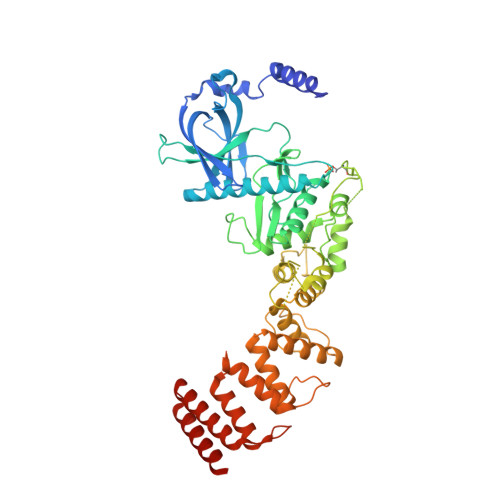
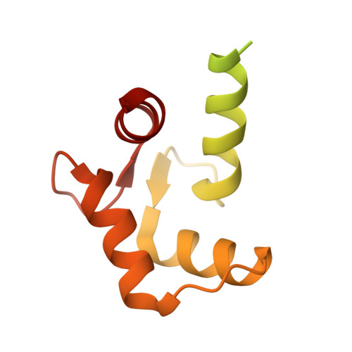
Structure of the complex between calmodulin and a functional construct of eukaryotic elongation factor 2 kinase bound to an ATP-competitive inhibitor.

Piserchio, A.Isiorho, E.A.Dalby, K.N.Ghose, R.

(2023) J Biol Chem 299: 104813-104813

  • DOI: https://doi.org/10.1016/j.jbc.2023.104813
  • Primary Citation of Related Structures:  
    8GM4, 8GM5

  • PubMed Abstract: 

    The calmodulin-activated α-kinase, eukaryotic elongation factor 2 kinase (eEF-2K), serves as a master regulator of translational elongation by specifically phosphorylating and reducing the ribosome affinity of the guanosine triphosphatase, eukaryotic elongation factor 2 (eEF-2). Given its critical role in a fundamental cellular process, dysregulation of eEF-2K has been implicated in several human diseases, including those of the cardiovascular system, chronic neuropathies, and many cancers, making it a critical pharmacological target. In the absence of high-resolution structural information, high-throughput screening efforts have yielded small-molecule candidates that show promise as eEF-2K antagonists. Principal among these is the ATP-competitive pyrido-pyrimidinedione inhibitor, A-484954, which shows high specificity toward eEF-2K relative to a panel of "typical" protein kinases. A-484954 has been shown to have some degree of efficacy in animal models of several disease states. It has also been widely deployed as a reagent in eEF-2K-specific biochemical and cell-biological studies. However, given the absence of structural information, the precise mechanism of the A-484954-mediated inhibition of eEF-2K has remained obscure. Leveraging our identification of the calmodulin-activatable catalytic core of eEF-2K, and our recent determination of its long-elusive structure, here we present the structural basis for its specific inhibition by A-484954. This structure, which represents the first for an inhibitor-bound catalytic domain of a member of the α-kinase family, enables rationalization of the existing structure-activity relationship data for A-484954 variants and lays the groundwork for further optimization of this scaffold to attain enhanced specificity/potency against eEF-2K.


  • Organizational Affiliation

    Department of Chemistry and Biochemistry, The City College of New York, New York, New York, USA.


Macromolecules
Find similar proteins by:  (by identity cutoff)  |  3D Structure
Entity ID: 1
MoleculeChains Sequence LengthOrganismDetailsImage
Eukaryotic elongation factor 2 kinase531Homo sapiensMutation(s): 0 
Gene Names: EEF2K
EC: 2.7.11.20
UniProt & NIH Common Fund Data Resources
Find proteins for O00418 (Homo sapiens)
Explore O00418 
Go to UniProtKB:  O00418
PHAROS:  O00418
GTEx:  ENSG00000103319 
Entity Groups  
Sequence Clusters30% Identity50% Identity70% Identity90% Identity95% Identity100% Identity
UniProt GroupO00418
Sequence Annotations
Expand
  • Reference Sequence
Find similar proteins by:  (by identity cutoff)  |  3D Structure
Entity ID: 2
MoleculeChains Sequence LengthOrganismDetailsImage
Calmodulin-1148Homo sapiensMutation(s): 0 
Gene Names: CALM1CALMCAMCAM1
UniProt & NIH Common Fund Data Resources
Find proteins for P0DP23 (Homo sapiens)
Explore P0DP23 
Go to UniProtKB:  P0DP23
PHAROS:  P0DP23
GTEx:  ENSG00000198668 
Entity Groups  
Sequence Clusters30% Identity50% Identity70% Identity90% Identity95% Identity100% Identity
UniProt GroupP0DP23
Sequence Annotations
Expand
  • Reference Sequence
Small Molecules
Ligands 4 Unique
IDChains Name / Formula / InChI Key2D Diagram3D Interactions
ADP
Query on ADP

Download Ideal Coordinates CCD File 
C [auth A]ADENOSINE-5'-DIPHOSPHATE
C10 H15 N5 O10 P2
XTWYTFMLZFPYCI-KQYNXXCUSA-N
EKI
Query on EKI

Download Ideal Coordinates CCD File 
D [auth A]7-amino-1-cyclopropyl-3-ethyl-2,4-dioxo-1,2,3,4-tetrahydropyrido[2,3-d]pyrimidine-6-carboxamide
C13 H15 N5 O3
HWODCHXORCTEGU-UHFFFAOYSA-N
ZN
Query on ZN

Download Ideal Coordinates CCD File 
E [auth A]ZINC ION
Zn
PTFCDOFLOPIGGS-UHFFFAOYSA-N
CA
Query on CA

Download Ideal Coordinates CCD File 
F [auth B],
G [auth B]
CALCIUM ION
Ca
BHPQYMZQTOCNFJ-UHFFFAOYSA-N
Modified Residues  1 Unique
IDChains TypeFormula2D DiagramParent
TPO
Query on TPO
A
L-PEPTIDE LINKINGC4 H10 N O6 PTHR
Experimental Data & Validation

Experimental Data

  • Method: X-RAY DIFFRACTION
  • Resolution: 2.12 Å
  • R-Value Free: 0.225 
  • R-Value Work: 0.181 
  • R-Value Observed: 0.183 
  • Space Group: P 1 21 1
Unit Cell:
Length ( Å )Angle ( ˚ )
a = 80.909α = 90
b = 61.041β = 111.26
c = 88.958γ = 90
Software Package:
Software NamePurpose
PHENIXrefinement
autoPROCdata processing
XDSdata reduction
Aimlessdata scaling
PHASERphasing
Cootmodel building

Structure Validation

View Full Validation Report


Ligand Structure Quality Assessment 


Entry History & Funding Information

Deposition Data


Funding OrganizationLocationGrant Number
National Institutes of Health/National Institute of General Medical Sciences (NIH/NIGMS)United StatesGM123252

Revision History  (Full details and data files)

  • Version 1.0: 2023-05-31
    Type: Initial release
  • Version 1.1: 2023-06-21
    Changes: Database references
  • Version 1.2: 2023-10-25
    Changes: Data collection