Pentaphosphorylation via the Anhydride of Dihydrogen Pentametaphosphate: Access to Nucleoside Hexa- and Heptaphosphates and Study of Their Interaction with Ribonuclease A.
Park, G., Wralstad, E.C., Faginas-Lago, N., Qian, K., Raines, R.T., Bistoni, G., Cummins, C.C.(2024) ACS Cent Sci 10: 1415-1422
- PubMed: 39071052 Search on PubMedSearch on PubMed Central
- DOI: https://doi.org/10.1021/acscentsci.4c00835
- Primary Citation Related Structures:
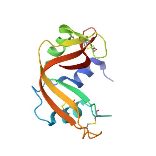
8FHM, 8GC9, 8GGG, 8S96 - PubMed Abstract:
Pentametaphosphate is the little studied cyclic pentamer of the metaphosphate ion, [PO 3 ] 5 5- . We show that the doubly protonated form of this pentamer can be selectively dehydrated to provide the anhydride [P 5 O 14 ] 3- ( 1 ). This trianion is the well-defined condensed phosphate component of a novel reagent for attachment of a pentaphosphate chain to biomolecules all in one go. Here, we demonstrate by extending adenosine monophosphate (AMP) and uridine monophosphate (UMP) to their corresponding nucleoside hexaphosphates, while adenosine diphosphate (ADP) and uridine diphosphate (UDP) are phosphate chain-extended to the corresponding nucleoside heptaphosphates. Such constructs are of interest for their potential biological function with respect to RNA-processing enzymes. Thus, we go on to investigate in detail the interaction of the polyanionic constructs with ribonuclease A, a model protein containing a polycationic active site and for which X-ray crystal structures are relatively straightforward to obtain. This work presents a combined experimental and quantum chemical approach to understanding the interactions of RNase A with the new nucleoside hexa- and heptaphosphate constructs.
- Department of Chemistry, Massachusetts Institute of Technology, Cambridge, Massachusetts 02139, United States.
Organizational Affiliation: