8GD7 | pdb_00008gd7

Loop Deleted DNA Polymerase Theta Polymerase Domain in Complex with Double Strand DNA Overhang and Inhibitor


Experimental Data Snapshot

  • Method: X-RAY DIFFRACTION
  • Resolution: 3.24 Å
  • R-Value Free: 
    0.244 (Depositor), 0.260 (DCC) 
  • R-Value Work: 
    0.228 (Depositor) 
  • R-Value Observed: 
    0.230 (Depositor) 

Starting Model: experimental
View more details

wwPDB Validation 3D Report Full Report

Validation slider image for 8GD7

Ligand Structure Quality Assessment 


This is version 1.0 of the entry. See complete history

Literature

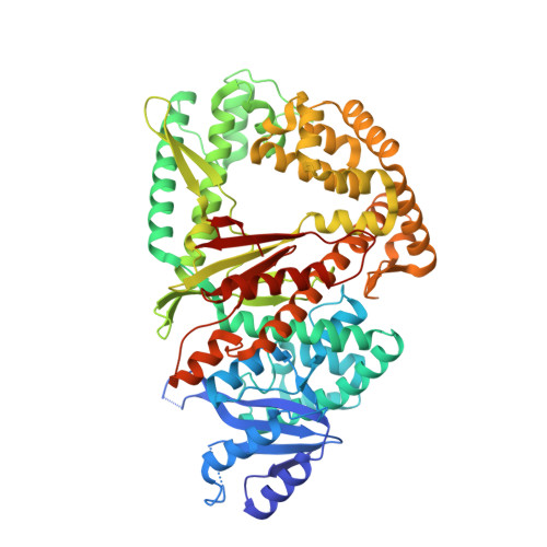
Discovery of a small-molecule inhibitor that traps Pol theta on DNA and synergizes with PARP inhibitors.

Fried, W.Tyagi, M.Minakhin, L.Chandramouly, G.Tredinnick, T.Ramanjulu, M.Auerbacher, W.Calbert, M.Rusanov, T.Hoang, T.Borisonnik, N.Betsch, R.Krais, J.J.Wang, Y.Vekariya, U.M.Gordon, J.Morton, G.Kent, T.Skorski, T.Johnson, N.Childers, W.Chen, X.S.Pomerantz, R.T.

(2024) Nat Commun 15: 2862-2862

  • DOI: https://doi.org/10.1038/s41467-024-46593-1
  • Primary Citation Related Structures: 
    8GD7

  • PubMed Abstract: 

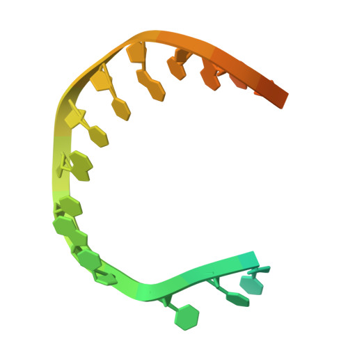
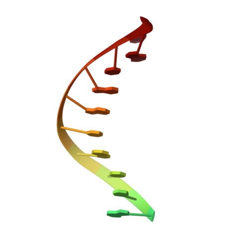
    The DNA damage response (DDR) protein DNA Polymerase θ (Polθ) is synthetic lethal with homologous recombination (HR) factors and is therefore a promising drug target in BRCA1/2 mutant cancers. We discover an allosteric Polθ inhibitor (Polθi) class with 4-6 nM IC 50 that selectively kills HR-deficient cells and acts synergistically with PARP inhibitors (PARPi) in multiple genetic backgrounds. X-ray crystallography and biochemistry reveal that Polθi selectively inhibits Polθ polymerase (Polθ-pol) in the closed conformation on B-form DNA/DNA via an induced fit mechanism. In contrast, Polθi fails to inhibit Polθ-pol catalytic activity on A-form DNA/RNA in which the enzyme binds in the open configuration. Remarkably, Polθi binding to the Polθ-pol:DNA/DNA closed complex traps the polymerase on DNA for more than forty minutes which elucidates the inhibitory mechanism of action. These data reveal a unique small-molecule DNA polymerase:DNA trapping mechanism that induces synthetic lethality in HR-deficient cells and potentiates the activity of PARPi.


  • Organizational Affiliation
    • Molecular and Computational Biology, Department of Biological Sciences and Chemistry, University of Southern California, Los Angeles, CA, USA.

Macromolecule Content 

  • Total Structure Weight: 85.22 kDa 
  • Atom Count: 5,514 
  • Modeled Residue Count: 654 
  • Deposited Residue Count: 690 
  • Unique protein chains: 1
  • Unique nucleic acid chains: 2

Macromolecules


Find similar proteins by:|  3D Structure
Entity ID: 1
MoleculeChains  Sequence LengthOrganismDetailsImage
DNA polymerase theta652Homo sapiensMutation(s): 0 
Gene Names: POLQ
EC: 2.7.7.7 (PDB Primary Data), 3.6.4.12 (UniProt)
UniProt & NIH Common Fund Data Resources
Find proteins for O75417 (Homo sapiens)
Explore O75417 
Go to UniProtKB:  O75417
PHAROS:  O75417
GTEx:  ENSG00000051341 
Entity Groups
Sequence Clusters30% Identity50% Identity70% Identity90% Identity95% Identity100% Identity
UniProt GroupO75417
Sequence Annotations
Expand
Reference Sequence
Find similar nucleic acids by:  Sequence
Entity ID: 2
MoleculeChains LengthOrganismImage
DNA TemplateB [auth E]24synthetic construct
Sequence Annotations
Expand
Reference Sequence
Find similar nucleic acids by:  Sequence
Entity ID: 3
MoleculeChains LengthOrganismImage
DNA PrimerC [auth F]14synthetic construct
Sequence Annotations
Expand
Reference Sequence

Small Molecules

Ligands 3 Unique
IDChains Name / Formula / InChI Key2D Diagram3D Interactions
DG3

Query on DG3


Download:Ideal Coordinates CCD File
E [auth A]2'-3'-DIDEOXYGUANOSINE-5'-TRIPHOSPHATE
C10 H16 N5 O12 P3
HDRRAMINWIWTNU-NTSWFWBYSA-N
Z5X
(Subject of Investigation/LOI)

Query on Z5X


Download:Ideal Coordinates CCD File
F [auth A]2-(3-methyl-2-oxoimidazolidin-1-yl)-4,6-bis(trifluoromethyl)phenyl (4-fluorophenyl)methylcarbamate
C20 H16 F7 N3 O3
OMLNQKKHSWHSNR-UHFFFAOYSA-N
MG

Query on MG


Download:Ideal Coordinates CCD File
D [auth A]MAGNESIUM ION
Mg
JLVVSXFLKOJNIY-UHFFFAOYSA-N

Experimental Data & Validation

Experimental Data

  • Method: X-RAY DIFFRACTION
  • Resolution: 3.24 Å
  • R-Value Free:  0.244 (Depositor), 0.260 (DCC) 
  • R-Value Work:  0.228 (Depositor) 
  • R-Value Observed: 0.230 (Depositor) 
Space Group: P 31 2 1
Unit Cell:
Length ( Å )Angle ( ˚ )
a = 171.433α = 90
b = 171.433β = 90
c = 63.167γ = 120
Software Package:
Software NamePurpose
PHENIXrefinement
PHASERphasing
Cootmodel building
HKL-2000data scaling
HKL-2000data reduction
JBluIce-EPICSdata collection

Structure Validation

View Full Validation Report


Ligand Structure Quality Assessment 


Entry History 

& Funding Information

Deposition Data


Funding OrganizationLocationGrant Number
National Institutes of Health/National Institute of General Medical Sciences (NIH/NIGMS)United StatesR01 GM130889

Revision History  (Full details and data files)

  • Version 1.0: 2024-06-19
    Type: Initial release