8GBS | pdb_00008gbs

Integrative model of the native Ana GV shell


Experimental Data Snapshot

  • Method: ELECTRON MICROSCOPY
  • Resolution: 8.00 Å
  • Aggregation State: HELICAL ARRAY 
  • Reconstruction Method: TOMOGRAPHY 

Starting Model: experimental
View more details

wwPDB Validation 3D Report Full Report

Validation slider image for 8GBS

This is version 1.3 of the entry. See complete history

Literature

Structure of Anabaena flos-aquae gas vesicles revealed by cryo-ET.

Dutka, P.Metskas, L.A.Hurt, R.C.Salahshoor, H.Wang, T.Y.Malounda, D.Lu, G.J.Chou, T.F.Shapiro, M.G.Jensen, G.J.

(2023) Structure 31: 518-528.e6

  • DOI: https://doi.org/10.1016/j.str.2023.03.011
  • Primary Citation Related Structures: 
    8GBS

  • PubMed Abstract: 

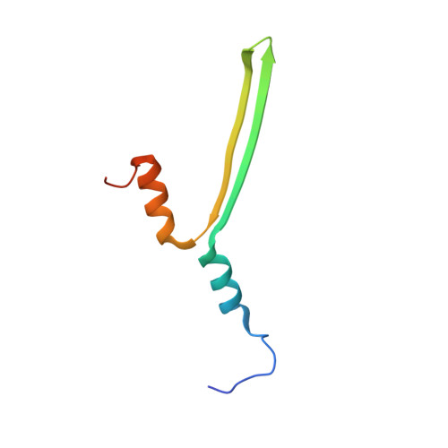
    Gas vesicles (GVs) are gas-filled protein nanostructures employed by several species of bacteria and archaea as flotation devices to enable access to optimal light and nutrients. The unique physical properties of GVs have led to their use as genetically encodable contrast agents for ultrasound and MRI. Currently, however, the structure and assembly mechanism of GVs remain unknown. Here we employ cryoelectron tomography to reveal how the GV shell is formed by a helical filament of highly conserved GvpA subunits. This filament changes polarity at the center of the GV cylinder, a site that may act as an elongation center. Subtomogram averaging reveals a corrugated pattern of the shell arising from polymerization of GvpA into a β sheet. The accessory protein GvpC forms a helical cage around the GvpA shell, providing structural reinforcement. Together, our results help explain the remarkable mechanical properties of GVs and their ability to adopt different diameters and shapes.


  • Organizational Affiliation
    • Division of Chemistry and Chemical Engineering, California Institute of Technology, Pasadena, CA 91125, USA; Division of Biology and Biological Engineering, California Institute of Technology, Pasadena, CA 91125, USA.

Macromolecule Content 

  • Total Structure Weight: 32.97 kDa 
  • Atom Count: 2,121 
  • Modeled Residue Count: 293 
  • Deposited Residue Count: 317 
  • Unique protein chains: 2

Macromolecules

Find similar proteins by:|  3D Structure
Entity ID: 1
MoleculeChains  Sequence LengthOrganismDetailsImage
Gas vesicle structural proteinA [auth A1],
B [auth A2],
C [auth A4],
E [auth A3]
71Dolichospermum flos-aquaeMutation(s): 0 
UniProt
Find proteins for P10397 (Dolichospermum flosaquae)
Explore P10397 
Go to UniProtKB:  P10397
Entity Groups
Sequence Clusters30% Identity50% Identity70% Identity90% Identity95% Identity100% Identity
UniProt GroupP10397
Sequence Annotations
Expand
Reference Sequence
Find similar proteins by:|  3D Structure
Entity ID: 2
MoleculeChains  Sequence LengthOrganismDetailsImage
Gas vesicle protein CD [auth C]33Dolichospermum flos-aquaeMutation(s): 0 

Experimental Data & Validation

Experimental Data

  • Method: ELECTRON MICROSCOPY
  • Resolution: 8.00 Å
  • Aggregation State: HELICAL ARRAY 
  • Reconstruction Method: TOMOGRAPHY 
EM Software:
TaskSoftware PackageVersion
RECONSTRUCTIONRELION

Structure Validation

View Full Validation Report


Entry History 

& Funding Information

Deposition Data


Funding OrganizationLocationGrant Number
National Institutes of Health/National Institute Of Allergy and Infectious Diseases (NIH/NIAID)United StatesAI127401
National Institutes of Health/National Institute of Biomedical Imaging and Bioengineering (NIH/NIBIB)United StatesEB018975

Revision History  (Full details and data files)

  • Version 1.0: 2023-04-19
    Type: Initial release
  • Version 1.1: 2023-04-26
    Changes: Database references
  • Version 1.2: 2023-05-17
    Changes: Database references
  • Version 1.3: 2024-06-19
    Changes: Data collection, Refinement description