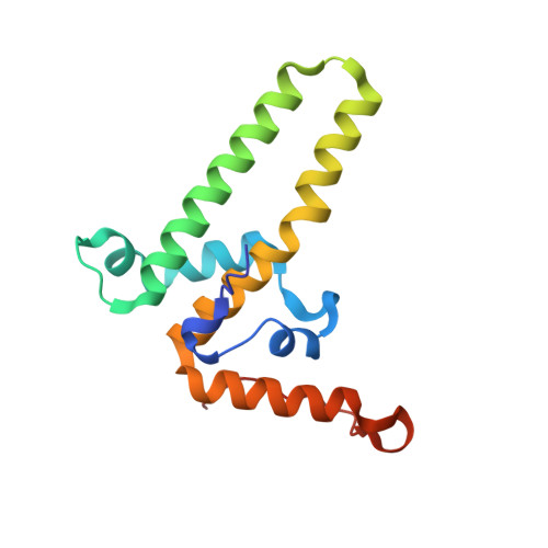
Structural basis for nuclear import of hepatitis B virus (HBV) nucleocapsid core.
Yang, R., Ko, Y.H., Li, F., Lokareddy, R.K., Hou, C.D., Kim, C., Klein, S., Antolinez, S., Marin, J.F., Perez-Segura, C., Jarrold, M.F., Zlotnick, A., Hadden-Perilla, J.A., Cingolani, G.(2024) Sci Adv 10: eadi7606-eadi7606
- PubMed: 38198557
- DOI: https://doi.org/10.1126/sciadv.adi7606
- Primary Citation of Related Structures:
7UMI, 8G5V, 8G6V, 8G8Y, 8GCN - PubMed Abstract:
Nuclear import of the hepatitis B virus (HBV) nucleocapsid is essential for replication that occurs in the nucleus. The ~360-angstrom HBV capsid translocates to the nuclear pore complex (NPC) as an intact particle, hijacking human importins in a reaction stimulated by host kinases. This paper describes the mechanisms of HBV capsid recognition by importins. We found that importin α1 binds a nuclear localization signal (NLS) at the far end of the HBV coat protein Cp183 carboxyl-terminal domain (CTD). This NLS is exposed to the capsid surface through a pore at the icosahedral quasi-sixfold vertex. Phosphorylation at serine-155, serine-162, and serine-170 promotes CTD compaction but does not affect the affinity for importin α1. The binding of 30 importin α1/β1 augments HBV capsid diameter to ~620 angstroms, close to the maximum size trafficable through the NPC. We propose that phosphorylation favors CTD externalization and prompts its compaction at the capsid surface, exposing the NLS to importins.
Organizational Affiliation:
Department of Biochemistry and Molecular Biology, Thomas Jefferson University, 1020 Locust Street, Philadelphia, PA 19107, USA.


