8G82 | pdb_00008g82

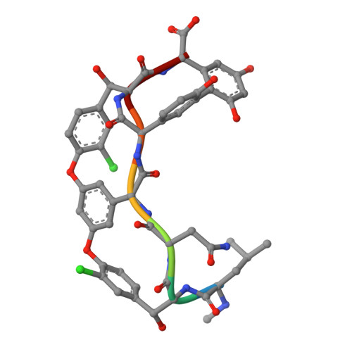
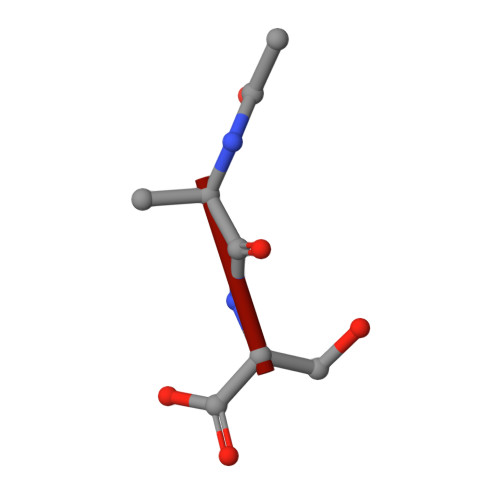
Vancomycin bound to D-Ala-D-Ser


Experimental Data Snapshot

  • Method: X-RAY DIFFRACTION
  • Resolution: 1.20 Å
  • R-Value Free: 
    0.171 (Depositor), 0.171 (DCC) 
  • R-Value Work: 
    0.153 (Depositor), 0.153 (DCC) 
  • R-Value Observed: 
    0.154 (Depositor) 

Starting Model: experimental
View more details

wwPDB Validation 3D Report Full Report

Validation slider image for 8G82

Ligand Structure Quality Assessment 


This is version 1.3 of the entry. See complete history

Literature

Crystal structure of vancomycin bound to the resistance determinant D-alanine-D-serine.

Park, J.H.Reviello, R.E.Loll, P.J.

(2024) IUCrJ 11: 133-139

  • DOI: https://doi.org/10.1107/S2052252524000289
  • Primary Citation Related Structures: 
    8G82

  • PubMed Abstract: 

    Vancomycin is a glycopeptide antibiotic that for decades has been a mainstay of treatment for persistent bacterial infections. However, the spread of antibiotic resistance threatens its continued utility. In particular, vancomycin-resistant enterococci (VRE) have become a pressing clinical challenge. Vancomycin acts by binding and sequestering the intermediate Lipid II in cell-wall biosynthesis, specifically recognizing a D-alanine-D-alanine dipeptide motif within the Lipid II molecule. VRE achieve resistance by remodeling this motif to either D-alanine-D-lactate or D-alanine-D-serine; the former substitution essentially abolishes recognition by vancomycin of Lipid II, whereas the latter reduces the affinity of the antibiotic by roughly one order of magnitude. The complex of vancomycin bound to D-alanine-D-serine has been crystallized, and its 1.20 Å X-ray crystal structure is presented here. This structure reveals that the D-alanine-D-serine ligand is bound in essentially the same position and same pose as the native D-alanine-D-alanine ligand. The serine-containing ligand appears to be slightly too large to be comfortably accommodated in this way, suggesting one possible contribution to the reduced binding affinity. In addition, two flexible hydroxyl groups - one from the serine side chain of the ligand, and the other from a glucose sugar on the antibiotic - are locked into single conformations in the complex, which is likely to contribute an unfavorable entropic component to the recognition of the serine-containing ligand.


  • Organizational Affiliation
    • Department of Biochemistry and Molecular Biology, Drexel University College of Medicine, PA 19102, USA.

Macromolecule Content 

  • Total Structure Weight: 17.44 kDa 
  • Atom Count: 1,542 
  • Modeled Residue Count: 100 
  • Deposited Residue Count: 100 
  • Unique protein chains: 2

Macromolecules

Find similar proteins by:  Sequence   |   3D Structure  
Entity ID: 1
MoleculeChains  Sequence LengthOrganismDetailsImage
Vancomycin
A, B, C, D, E
A, B, C, D, E, F, G, H, I, J
7Amycolatopsis orientalisMutation(s): 0 
Find similar proteins by:  Sequence   |   3D Structure  
Entity ID: 2
MoleculeChains  Sequence LengthOrganismDetailsImage
D-Ala-D-Ser
K, L, M, N, O
K, L, M, N, O, P, Q, R, S, T
3synthetic constructMutation(s): 0 

Oligosaccharides

Help  
Entity ID: 3
MoleculeChains Length2D Diagram GlycosylationD Interactions
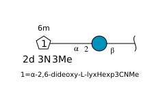
vancosamine-(1-2)-beta-D-glucopyranose
AA [auth k],
BA [auth m],
U,
V [auth W],
W [auth Y],
AA [auth k],
BA [auth m],
U,
V [auth W],
W [auth Y],
X [auth a],
Y [auth c],
Z [auth e]
2N/A
Glycosylation Resources
GlyTouCan: G14263HU
GlyCosmos: G14263HU

Small Molecules

Ligands 5 Unique
IDChains Name / Formula / InChI Key2D Diagram3D Interactions
BGC

Query on BGC


Download:Ideal Coordinates CCD File
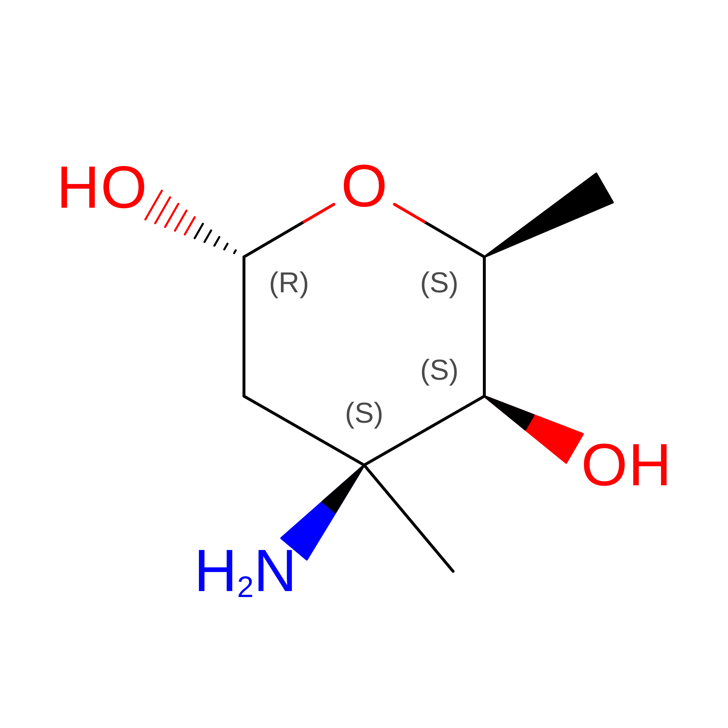
FA [auth G],
JA [auth H]
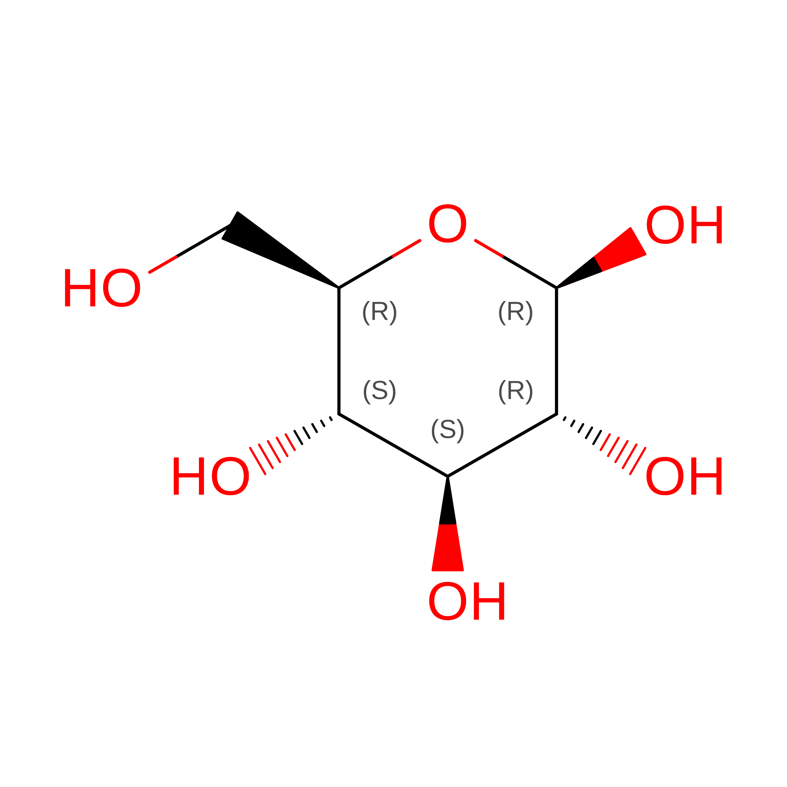
beta-D-glucopyranose
C6 H12 O6
WQZGKKKJIJFFOK-VFUOTHLCSA-N
RER

Query on RER


Download:Ideal Coordinates CCD File
GA [auth G],
KA [auth H]
vancosamine
C7 H15 N O3
OIJZDPGKNVKVBL-WNJXEPBRSA-N
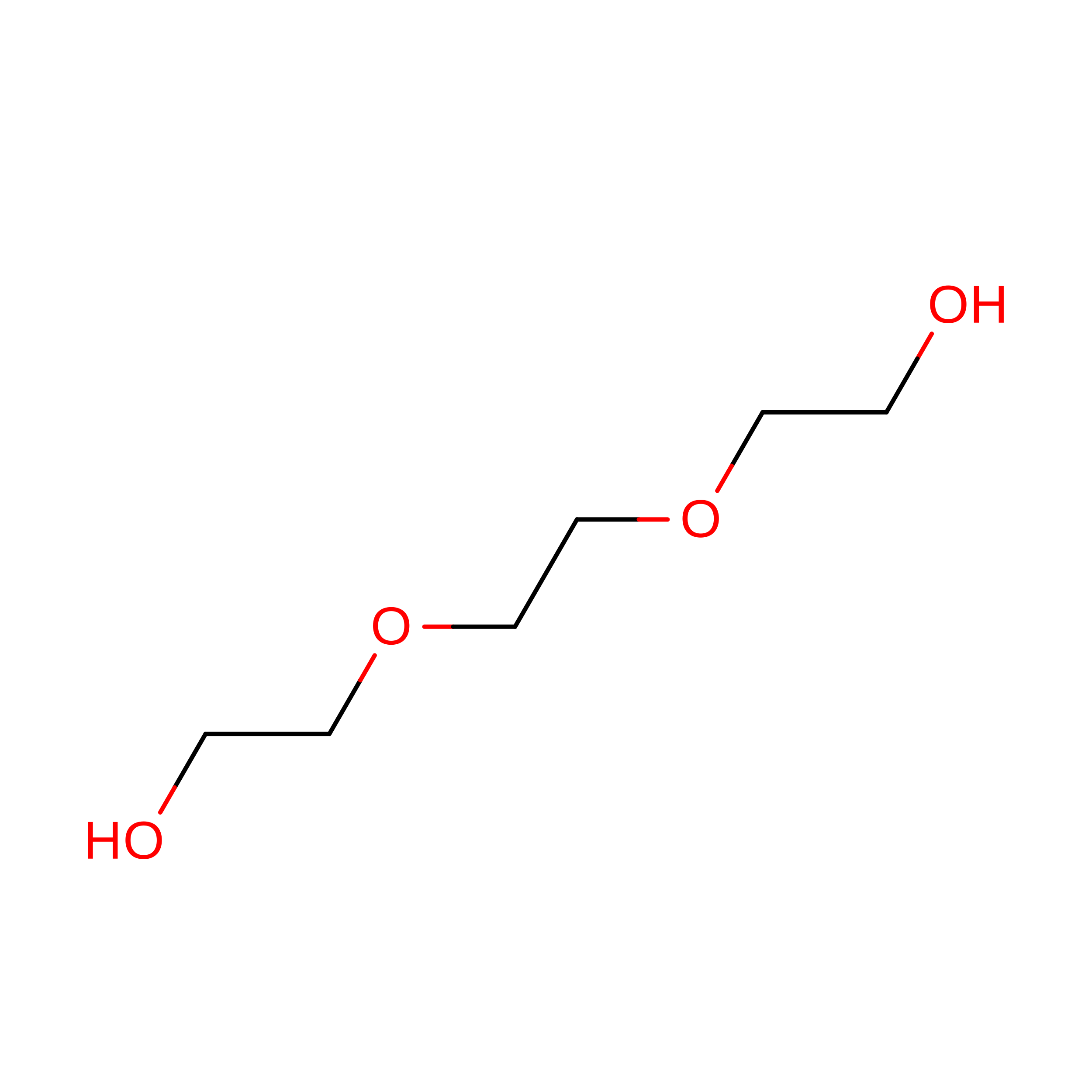
PGE

Query on PGE


Download:Ideal Coordinates CCD File
CA [auth A]TRIETHYLENE GLYCOL
C6 H14 O4
ZIBGPFATKBEMQZ-UHFFFAOYSA-N
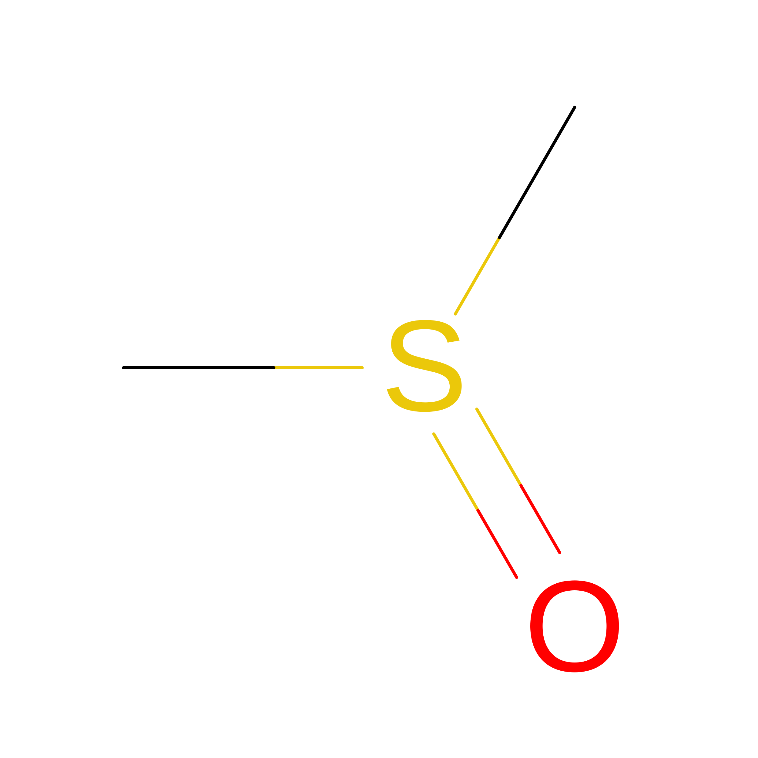
DMS

Query on DMS


Download:Ideal Coordinates CCD File
EA [auth G],
HA [auth H],
IA [auth H],
MA [auth J]
DIMETHYL SULFOXIDE
C2 H6 O S
IAZDPXIOMUYVGZ-UHFFFAOYSA-N
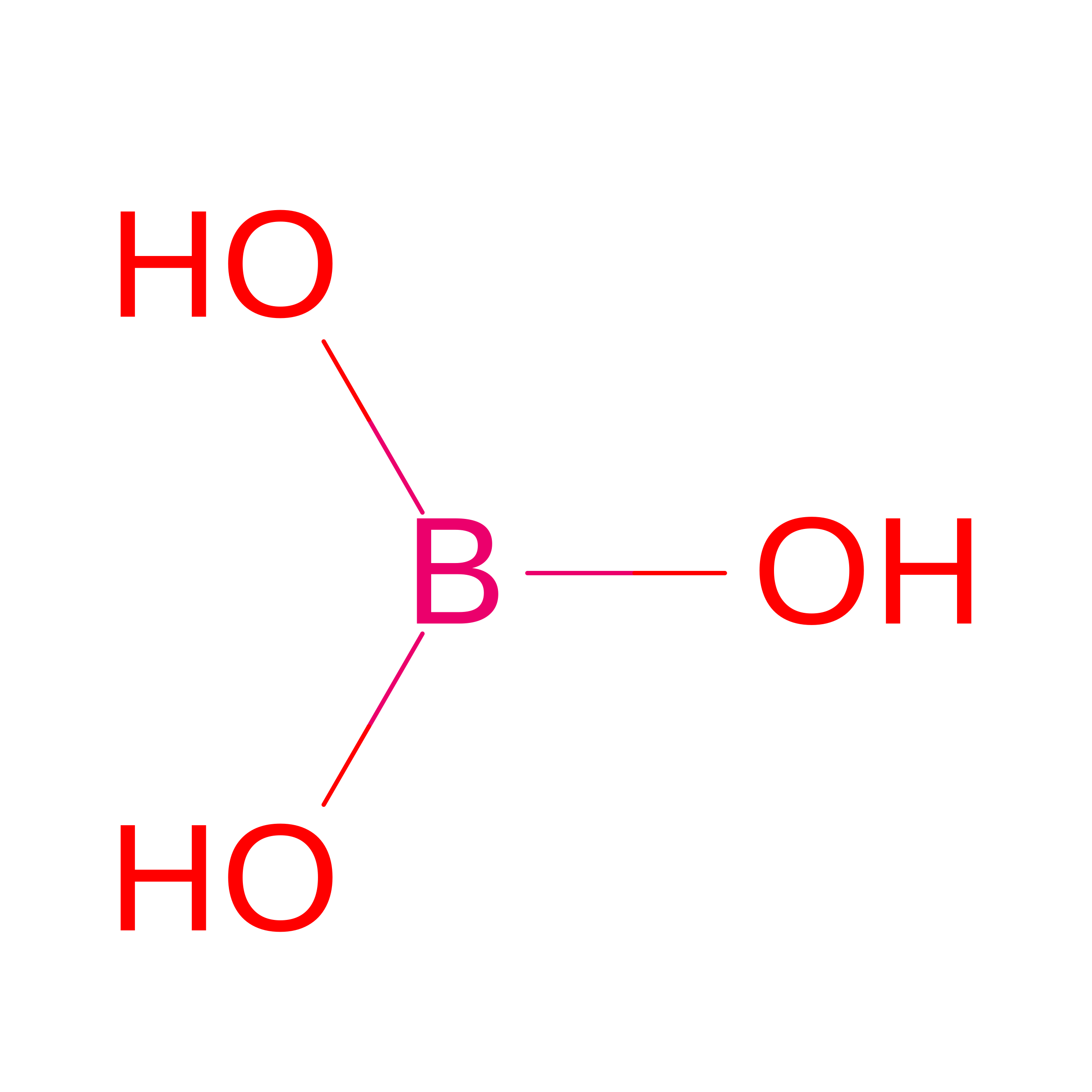
BO3

Query on BO3


Download:Ideal Coordinates CCD File
DA [auth D],
LA [auth J],
NA [auth O]
BORIC ACID
B H3 O3
KGBXLFKZBHKPEV-UHFFFAOYSA-N
Modified Residues  1 Unique
IDChains TypeFormula2D DiagramParent
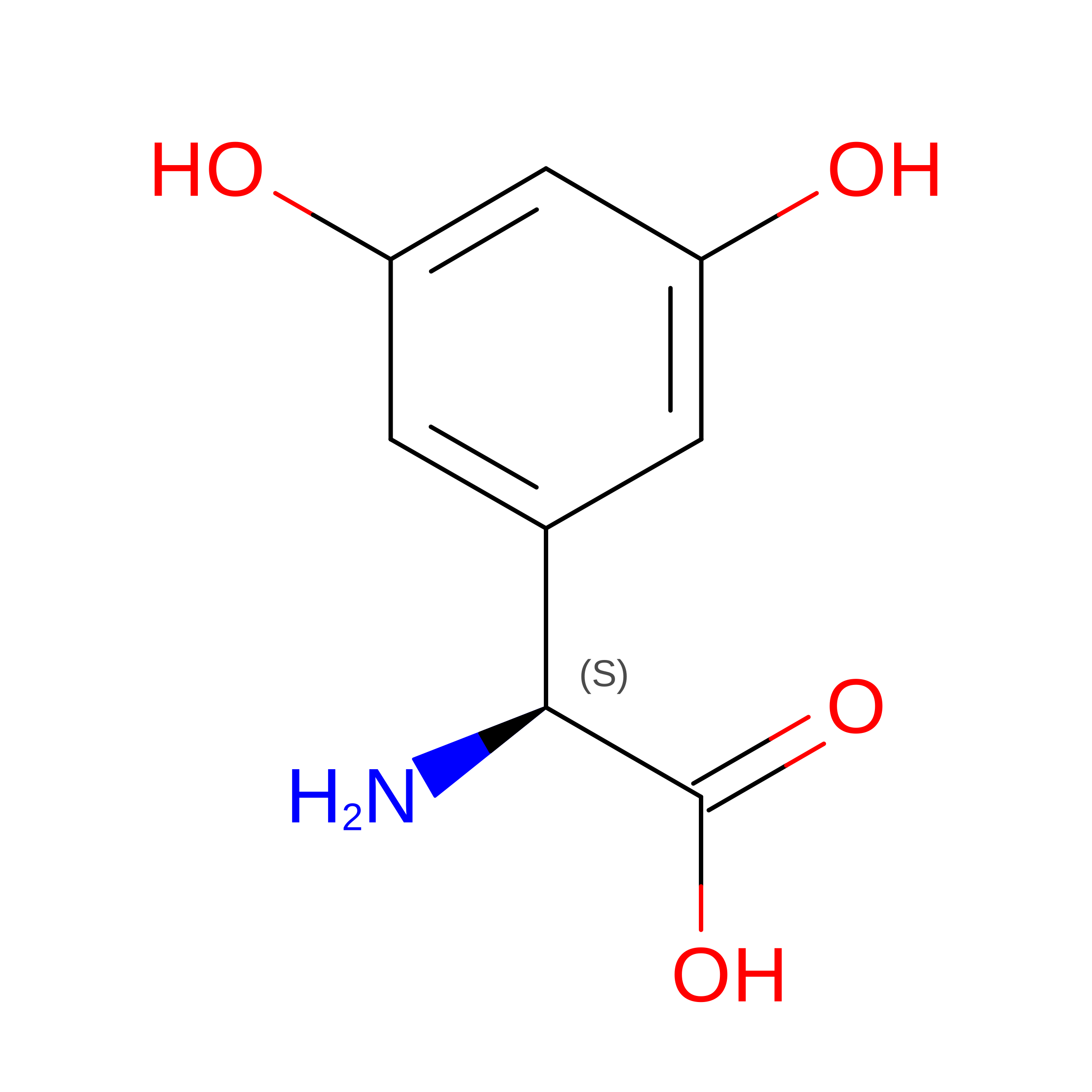
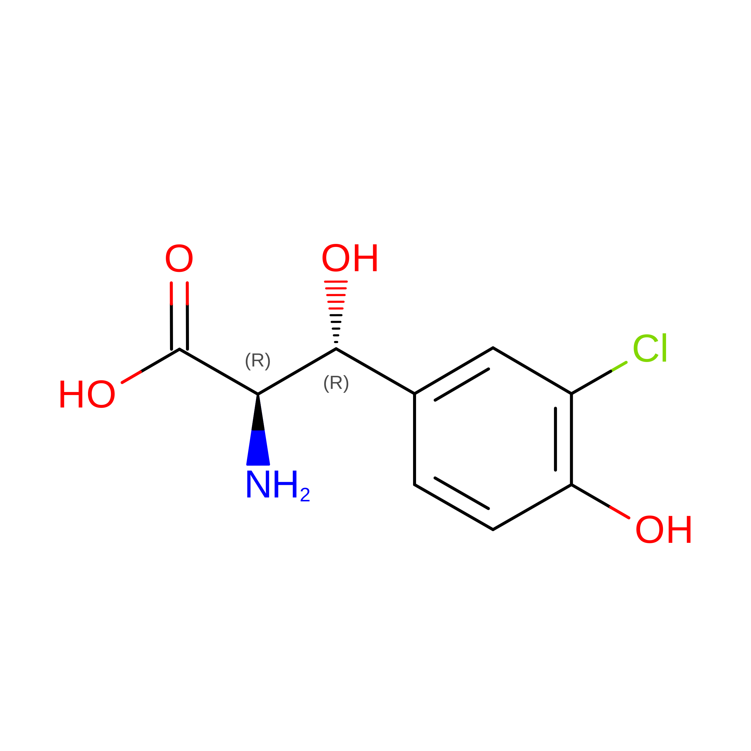
3FG
Query on 3FG
A, B, C, D, E
A, B, C, D, E, F, G, H, I, J
L-PEPTIDE LINKINGC8 H9 N O4

--

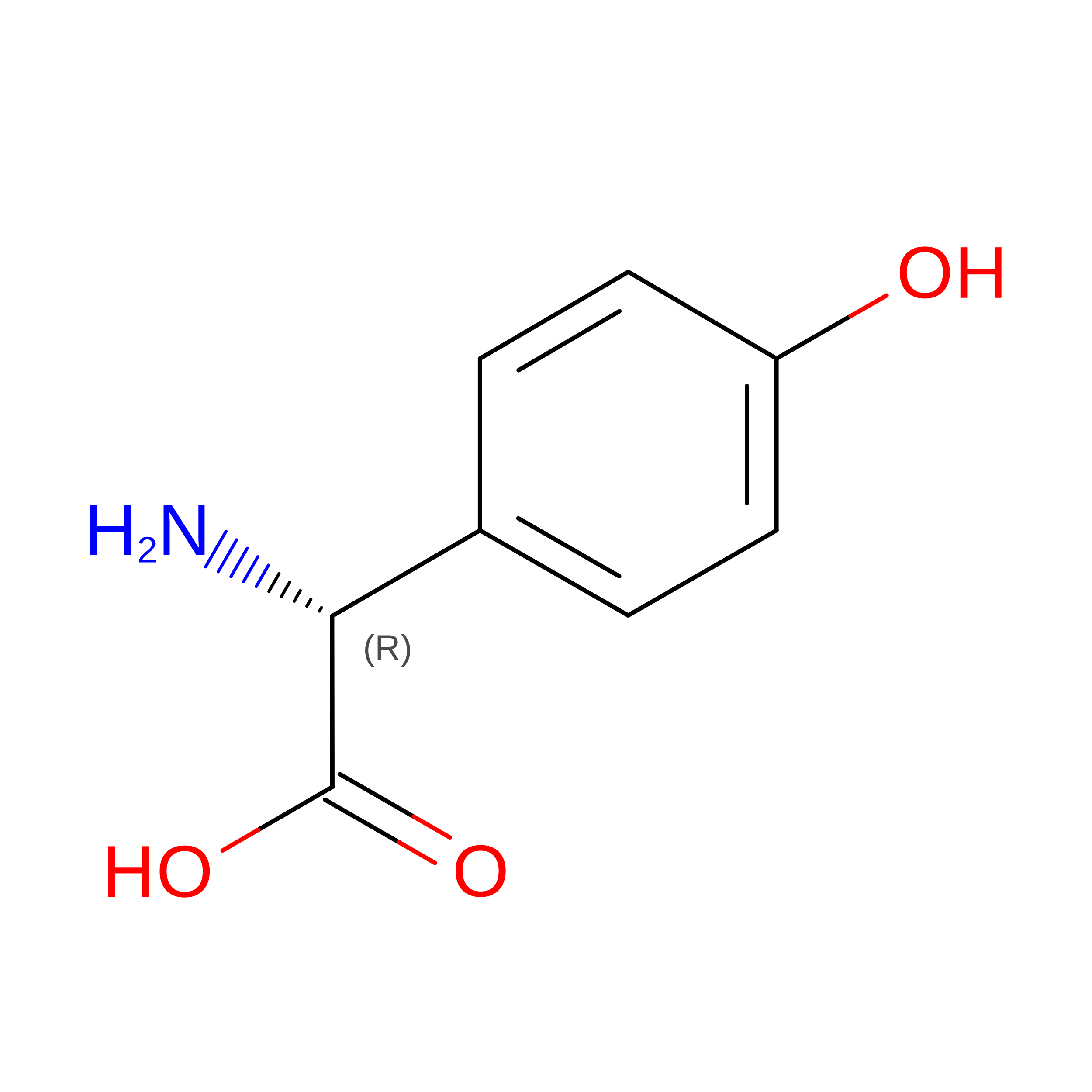
GHP
Query on GHP
A, B, C, D, E
A, B, C, D, E, F, G, H, I, J
D-PEPTIDE LINKINGC8 H9 N O3

--

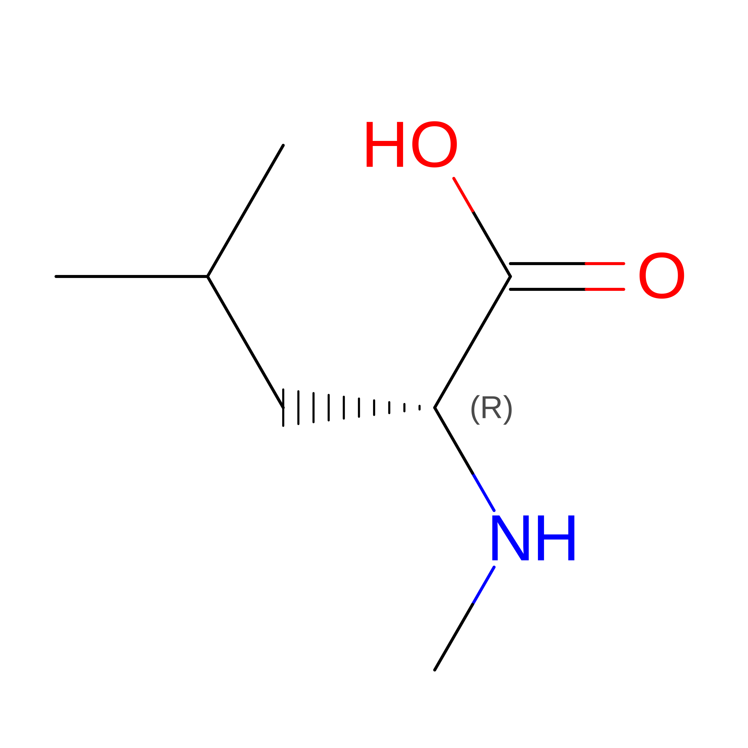
MLU
Query on MLU
A, B, C, D, E
A, B, C, D, E, F, G, H, I, J
D-PEPTIDE LINKINGC7 H15 N O2

--

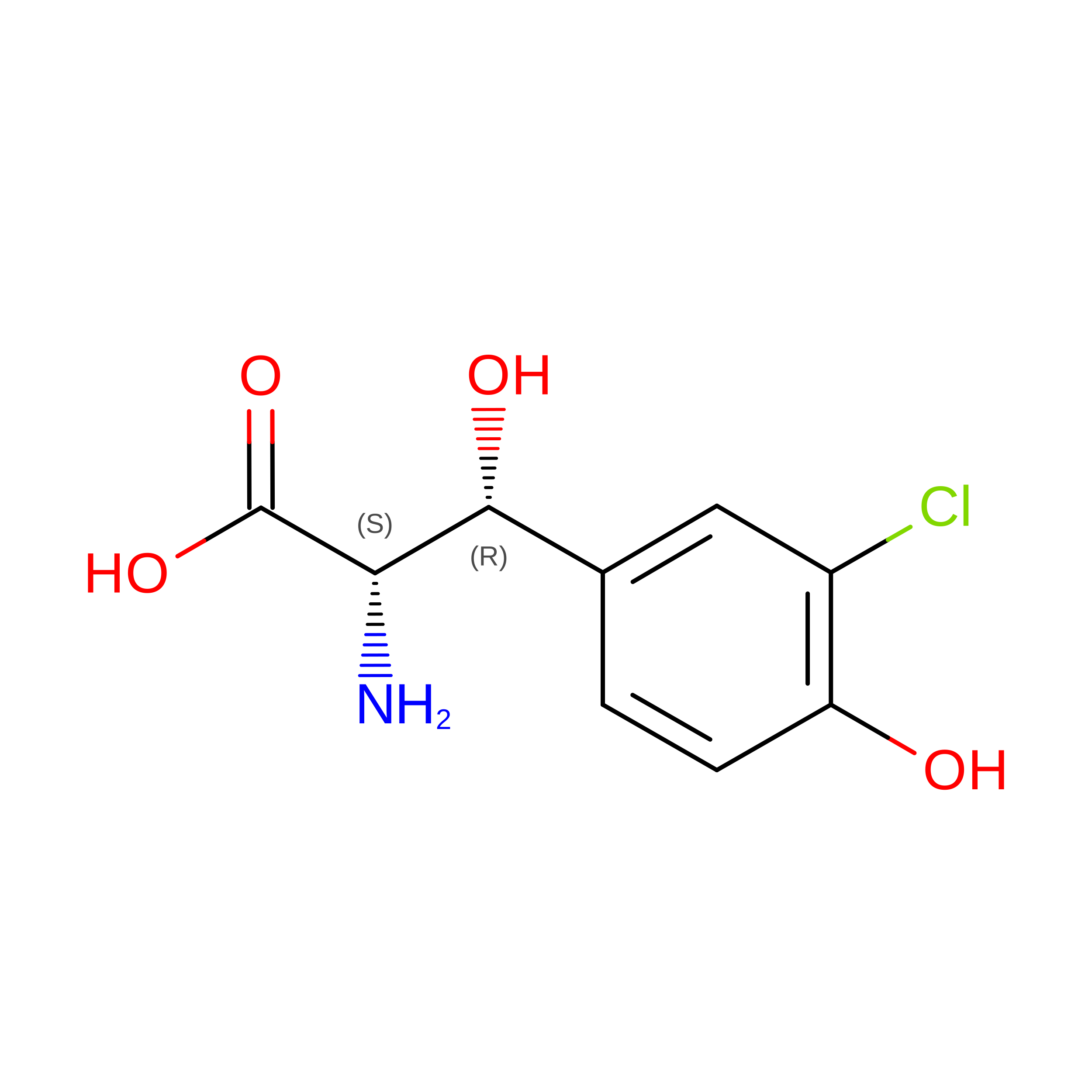
OMY
Query on OMY
A, B, C, D, E
A, B, C, D, E, F, G, H, I, J
L-PEPTIDE LINKINGC9 H10 Cl N O4TYR
OMZ
Query on OMZ
A, B, C, D, E
A, B, C, D, E, F, G, H, I, J
D-PEPTIDE LINKINGC9 H10 Cl N O4

--

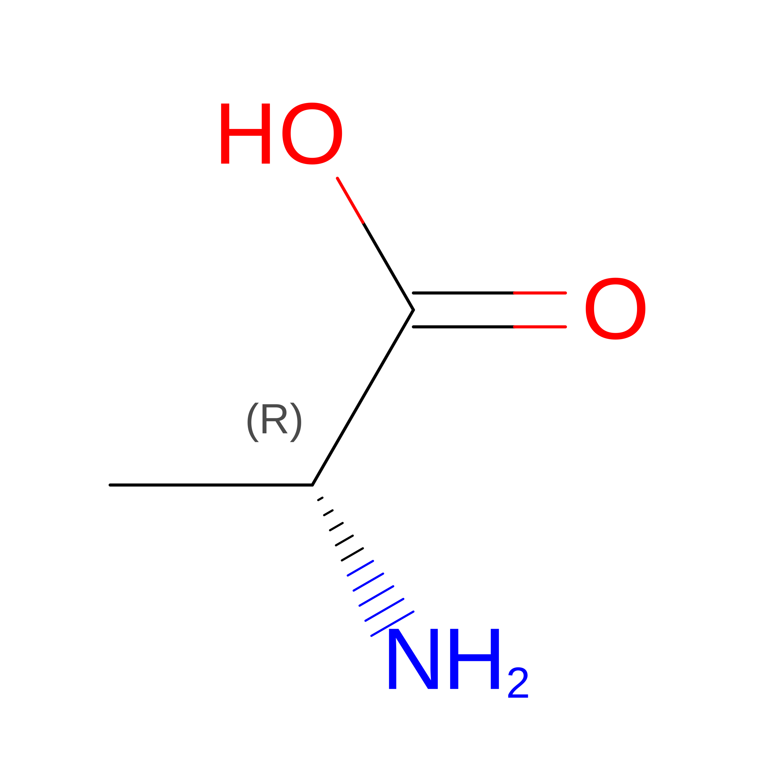
DAL
Query on DAL
K, L, M, N, O
K, L, M, N, O, P, Q, R, S, T
D-PEPTIDE LINKINGC3 H7 N O2

--

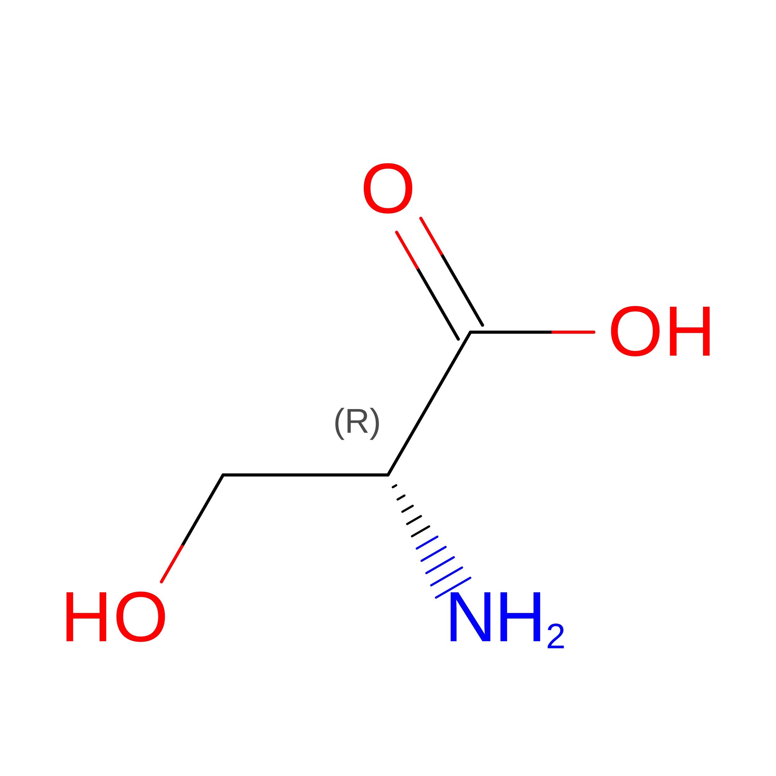
DSN
Query on DSN
K, L, M, N, O
K, L, M, N, O, P, Q, R, S, T
D-PEPTIDE LINKINGC3 H7 N O3

--

Experimental Data & Validation

Experimental Data

  • Method: X-RAY DIFFRACTION
  • Resolution: 1.20 Å
  • R-Value Free:  0.171 (Depositor), 0.171 (DCC) 
  • R-Value Work:  0.153 (Depositor), 0.153 (DCC) 
  • R-Value Observed: 0.154 (Depositor) 
Space Group: P 21 21 21
Unit Cell:
Length ( Å )Angle ( ˚ )
a = 30.49α = 90
b = 71.15β = 90
c = 82.44γ = 90
Software Package:
Software NamePurpose
PHENIXrefinement
XDSdata reduction
XSCALEdata scaling
PHENIXphasing

Structure Validation

View Full Validation Report


Ligand Structure Quality Assessment 


Entry History 

& Funding Information

Deposition Data


Funding OrganizationLocationGrant Number
National Institutes of Health/National Institute Of Allergy and Infectious Diseases (NIH/NIAID)United StatesR01AI148679

Revision History  (Full details and data files)

  • Version 1.0: 2024-01-24
    Type: Initial release
  • Version 1.1: 2024-02-07
    Changes: Database references
  • Version 1.2: 2024-03-13
    Changes: Database references
  • Version 1.3: 2024-03-20
    Changes: Database references