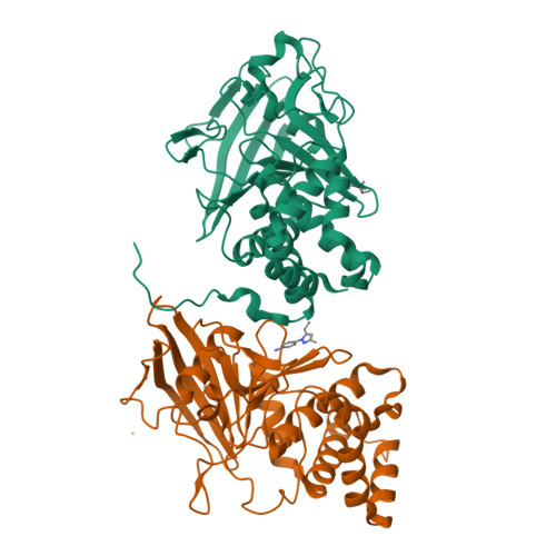
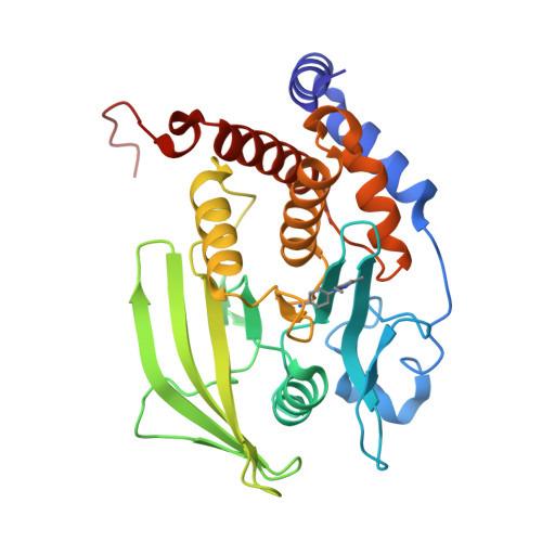
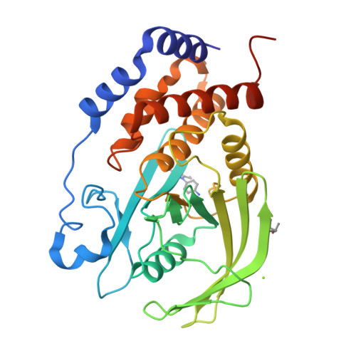
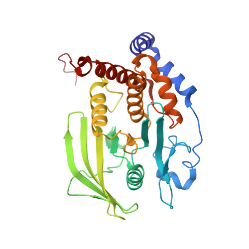
RCSB PDB - 8G69: Wildtype PTP1b in complex with DES5743

 8G69

Wildtype PTP1b in complex with DES5743


Experimental Data Snapshot

  • Method: X-RAY DIFFRACTION
  • Resolution: 1.53 Å
  • R-Value Free: 0.198 
  • R-Value Work: 0.169 
  • R-Value Observed: 0.171 

Starting Model: experimental
View more details

wwPDB Validation   3D Report Full Report


Ligand Structure Quality Assessment 

Created with Raphaël 2.3.0Worse 01 BetterLigand structure goodness of fit to experimental dataBest fitted YXNClick on this verticalbar to view details

This is version 1.3 of the entry. See complete history


Literature

Discovery and Validation of the Binding Poses of Allosteric Fragment Hits to Protein Tyrosine Phosphatase 1b: From Molecular Dynamics Simulations to X-ray Crystallography.

Greisman, J.B.Willmore, L.Yeh, C.Y.Giordanetto, F.Shahamadtar, S.Nisonoff, H.Maragakis, P.Shaw, D.E.

(2023) J Chem Inf Model 63: 2644-2650

  • DOI: https://doi.org/10.1021/acs.jcim.3c00236
  • Primary Citation of Related Structures:  
    8G65, 8G67, 8G68, 8G69, 8G6A

  • PubMed Abstract: 

    Fragment-based drug discovery has led to six approved drugs, but the small sizes of the chemical fragments used in such methods typically result in only weak interactions between the fragment and its target molecule, which makes it challenging to experimentally determine the three-dimensional poses fragments assume in the bound state. One computational approach that could help address this difficulty is long-timescale molecular dynamics (MD) simulations, which have been used in retrospective studies to recover experimentally known binding poses of fragments. Here, we present the results of long-timescale MD simulations that we used to prospectively discover binding poses for two series of fragments in allosteric pockets on a difficult and important pharmaceutical target, protein tyrosine phosphatase 1b (PTP1b). Our simulations reversibly sampled the fragment association and dissociation process. One of the binding pockets found in the simulations has not to our knowledge been previously observed with a bound fragment, and the other pocket adopted a very rare conformation. We subsequently obtained high-resolution crystal structures of members of each fragment series bound to PTP1b, and the experimentally observed poses confirmed the simulation results. To the best of our knowledge, our findings provide the first demonstration that MD simulations can be used prospectively to determine fragment binding poses to previously unidentified pockets.


  • Organizational Affiliation

    D. E. Shaw Research, New York, New York 10036, United States.


Macromolecules
Find similar proteins by:  (by identity cutoff)  |  3D Structure
Entity ID: 1
MoleculeChains Sequence LengthOrganismDetailsImage
Tyrosine-protein phosphatase non-receptor type 1
A, B
298Homo sapiensMutation(s): 0 
Gene Names: PTPN1PTP1B
EC: 3.1.3.48
UniProt & NIH Common Fund Data Resources
Find proteins for P18031 (Homo sapiens)
Explore P18031 
Go to UniProtKB:  P18031
PHAROS:  P18031
GTEx:  ENSG00000196396 
Entity Groups  
Sequence Clusters30% Identity50% Identity70% Identity90% Identity95% Identity100% Identity
UniProt GroupP18031
Sequence Annotations
Expand
  • Reference Sequence
Experimental Data & Validation

Experimental Data

  • Method: X-RAY DIFFRACTION
  • Resolution: 1.53 Å
  • R-Value Free: 0.198 
  • R-Value Work: 0.169 
  • R-Value Observed: 0.171 
  • Space Group: P 43 21 2
Unit Cell:
Length ( Å )Angle ( ˚ )
a = 88.07α = 90
b = 88.07β = 90
c = 162.36γ = 90
Software Package:
Software NamePurpose
REFMACrefinement
xia2data reduction
Aimlessdata scaling
PHASERphasing

Structure Validation

View Full Validation Report


Ligand Structure Quality Assessment 

Created with Raphaël 2.3.0Worse 01 BetterLigand structure goodness of fit to experimental dataBest fitted YXNClick on this verticalbar to view details

Entry History & Funding Information

Deposition Data


Funding OrganizationLocationGrant Number
Not funded--

Revision History  (Full details and data files)

  • Version 1.0: 2023-04-26
    Type: Initial release
  • Version 1.1: 2023-05-03
    Changes: Database references
  • Version 1.2: 2023-05-17
    Changes: Database references
  • Version 1.3: 2024-05-22
    Changes: Data collection