8G1E | pdb_00008g1e

Structure of ACLY-D1026A-products-asym

  • Classification: TRANSFERASE
  • Organism(s): Homo sapiens
  • Expression System: Escherichia coli
  • Mutation(s): Yes 

  • Deposited: 2023-02-02 Released: 2023-05-10 
  • Deposition Author(s): Wei, X., Marmorstein, R.
  • Funding Organization(s): National Institutes of Health/National Institute of General Medical Sciences (NIH/NIGMS)

Experimental Data Snapshot

  • Method: ELECTRON MICROSCOPY
  • Resolution: 2.80 Å
  • Aggregation State: PARTICLE 
  • Reconstruction Method: SINGLE PARTICLE 

wwPDB Validation 3D Report Full Report

Validation slider image for 8G1E

This is version 1.1 of the entry. See complete history

Literature

Allosteric role of the citrate synthase homology domain of ATP citrate lyase.

Wei, X.Schultz, K.Pepper, H.L.Megill, E.Vogt, A.Snyder, N.W.Marmorstein, R.

(2023) Nat Commun 14: 2247-2247

  • DOI: https://doi.org/10.1038/s41467-023-37986-9
  • Primary Citation Related Structures: 
    7RIG, 7RKZ, 7RMP, 8G1E, 8G1F, 8G5C, 8G5D

  • PubMed Abstract: 

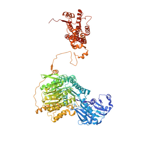
    ATP citrate lyase (ACLY) is the predominant nucleocytosolic source of acetyl-CoA and is aberrantly regulated in many diseases making it an attractive therapeutic target. Structural studies of ACLY reveal a central homotetrameric core citrate synthase homology (CSH) module flanked by acyl-CoA synthetase homology (ASH) domains, with ATP and citrate binding the ASH domain and CoA binding the ASH-CSH interface to produce acetyl-CoA and oxaloacetate products. The specific catalytic role of the CSH module and an essential D1026A residue contained within it has been a matter of debate. Here, we report biochemical and structural analysis of an ACLY-D1026A mutant demonstrating that this mutant traps a (3S)-citryl-CoA intermediate in the ASH domain in a configuration that is incompatible with the formation of acetyl-CoA, is able to convert acetyl-CoA and OAA to (3S)-citryl-CoA in the ASH domain, and can load CoA and unload acetyl-CoA in the CSH module. Together, this data support an allosteric role for the CSH module in ACLY catalysis.


  • Organizational Affiliation
    • Department of Biochemistry & Biophysics, Perelman School of Medicine, University of Pennsylvania, Philadelphia, PA, 19104, USA.

Macromolecule Content 

  • Total Structure Weight: 491.49 kDa 
  • Atom Count: 32,649 
  • Modeled Residue Count: 4,148 
  • Deposited Residue Count: 4,404 
  • Unique protein chains: 1

Macromolecules

Find similar proteins by:|  3D Structure
Entity ID: 1
MoleculeChains  Sequence LengthOrganismDetailsImage
ATP-citrate synthase
A, B, C, D
1,101Homo sapiensMutation(s): 1 
Gene Names: ACLY
EC: 2.3.3.8
UniProt & NIH Common Fund Data Resources
Find proteins for P53396 (Homo sapiens)
Explore P53396 
Go to UniProtKB:  P53396
PHAROS:  P53396
GTEx:  ENSG00000131473 
Entity Groups
Sequence Clusters30% Identity50% Identity70% Identity90% Identity95% Identity100% Identity
UniProt GroupP53396
Sequence Annotations
Expand
Reference Sequence

Small Molecules

Ligands 6 Unique
IDChains Name / Formula / InChI Key2D Diagram3D Interactions
Q5B

Query on Q5B


Download:Ideal Coordinates CCD File
I [auth B],
P [auth C],
T [auth D]
(3S)-citryl-Coenzyme A
C27 H42 N7 O22 P3 S
IHVFHZGGMJDGGZ-VPXVXCNZSA-N
ACO

Query on ACO


Download:Ideal Coordinates CCD File
E [auth A],
J [auth B],
L [auth C],
R [auth D]
ACETYL COENZYME *A
C23 H38 N7 O17 P3 S
ZSLZBFCDCINBPY-ZSJPKINUSA-N
ADP

Query on ADP


Download:Ideal Coordinates CCD File
N [auth C],
U [auth D]
ADENOSINE-5'-DIPHOSPHATE
C10 H15 N5 O10 P2
XTWYTFMLZFPYCI-KQYNXXCUSA-N
OAA

Query on OAA


Download:Ideal Coordinates CCD File
F [auth A],
G [auth A],
M [auth C],
S [auth D]
OXALOACETATE ION
C4 H3 O5
KHPXUQMNIQBQEV-UHFFFAOYSA-M
PO4

Query on PO4


Download:Ideal Coordinates CCD File
K [auth B],
Q [auth C],
V [auth D]
PHOSPHATE ION
O4 P
NBIIXXVUZAFLBC-UHFFFAOYSA-K
UNL(
Subject of Investigation/LOI)

Query on UNL


Download:Ideal Coordinates CCD File
H [auth B],
O [auth C]
Unknown ligand
NBIIXXVUZAFLBC-UHFFFAOYSA-K

Experimental Data & Validation

Experimental Data

  • Method: ELECTRON MICROSCOPY
  • Resolution: 2.80 Å
  • Aggregation State: PARTICLE 
  • Reconstruction Method: SINGLE PARTICLE 

Structure Validation

View Full Validation Report


Entry History 

& Funding Information

Deposition Data


Funding OrganizationLocationGrant Number
National Institutes of Health/National Institute of General Medical Sciences (NIH/NIGMS)United States--

Revision History  (Full details and data files)

  • Version 1.0: 2023-05-10
    Type: Initial release
  • Version 1.1: 2024-06-19
    Changes: Data collection