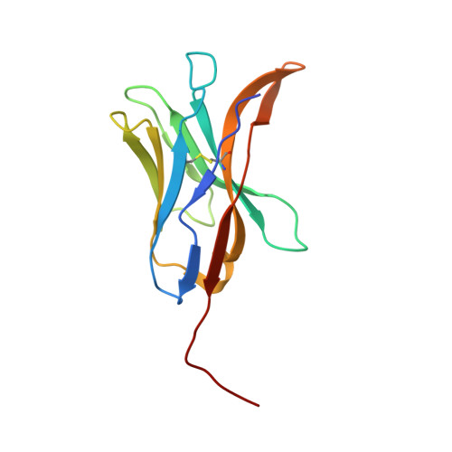
8FTT | pdb_00008ftt

Crystal structure of a glycosylase specific nanobody


Experimental Data Snapshot

  • Method: X-RAY DIFFRACTION
  • Resolution: 2.08 Å
  • R-Value Free: 
    0.229 (Depositor), 0.236 (DCC) 
  • R-Value Work: 
    0.199 (Depositor), 0.209 (DCC) 
  • R-Value Observed: 
    0.252 (Depositor) 

Starting Model: experimental
View more details

wwPDB Validation 3D Report Full Report

Validation slider image for 8FTT

This is version 1.2 of the entry. See complete history

Literature

Deciphering the crystal structure of a novel nanobody against the NEIL1 DNA glycosylase.

Thompson, M.K.Sharma, N.Thorn, A.Prakash, A.

(2024) Acta Crystallogr D Struct Biol 80: 137-146

  • DOI: https://doi.org/10.1107/S205979832400038X
  • Primary Citation Related Structures: 
    8FTT

  • PubMed Abstract: 

    Nanobodies (VHHs) are single-domain antibodies with three antigenic CDR regions and are used in diverse scientific applications. Here, an ∼14 kDa nanobody (A5) specific for the endonuclease VIII (Nei)-like 1 or NEIL1 DNA glycosylase involved in the first step of the base-excision repair pathway was crystallized and its structure was determined to 2.1 Å resolution. The crystals posed challenges due to potential twinning and anisotropic diffraction. Despite inconclusive twinning indicators, reprocessing in an orthorhombic setting and molecular replacement in space group P2 1 2 1 2 enabled the successful modeling of 96% of residues in the asymmetric unit, with final R work and R free values of 0.199 and 0.229, respectively.


  • Organizational Affiliation
    • Mitchell Cancer Institute, University of South Alabama Health, 1660 Springhill Avenue, Mobile, AL 36604, USA.

Macromolecule Content 

  • Total Structure Weight: 59.26 kDa 
  • Atom Count: 3,995 
  • Modeled Residue Count: 501 
  • Deposited Residue Count: 520 
  • Unique protein chains: 1

Macromolecules

Find similar proteins by:|  3D Structure
Entity ID: 1
MoleculeChains  Sequence LengthOrganismDetailsImage
Anti-NEIL1 Nanobody
A, B, C, D
130synthetic constructMutation(s): 0 

Experimental Data & Validation

Experimental Data

  • Method: X-RAY DIFFRACTION
  • Resolution: 2.08 Å
  • R-Value Free:  0.229 (Depositor), 0.236 (DCC) 
  • R-Value Work:  0.199 (Depositor), 0.209 (DCC) 
  • R-Value Observed: 0.252 (Depositor) 
Space Group: P 21 21 2
Unit Cell:
Length ( Å )Angle ( ˚ )
a = 121.806α = 90
b = 76.022β = 90
c = 75.767γ = 90
Software Package:
Software NamePurpose
PHENIXrefinement
Cootmodel building
SAINTdata reduction
Aimlessdata scaling
PHASERphasing

Structure Validation

View Full Validation Report


Entry History 

& Funding Information

Deposition Data


Funding OrganizationLocationGrant Number
National Institutes of Health/National Institute of Environmental Health Sciences (NIH/NIEHS)United StatesONES R01-ES030084

Revision History  (Full details and data files)

  • Version 1.0: 2023-10-11
    Type: Initial release
  • Version 1.1: 2024-11-13
    Changes: Structure summary
  • Version 1.2: 2025-03-19
    Changes: Database references