RCSB PDB - 8FS2: CamA Adenine Methyltransferase Complexed to Cognate Substrate DNA and Inhibitor 11b (YD907)

 8FS2

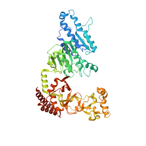
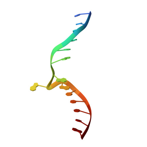
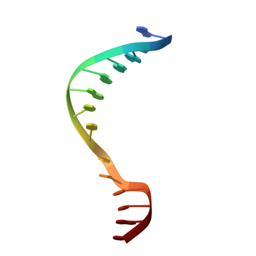
CamA Adenine Methyltransferase Complexed to Cognate Substrate DNA and Inhibitor 11b (YD907)


Experimental Data Snapshot

  • Method: X-RAY DIFFRACTION
  • Resolution: 2.59 Å
  • R-Value Free: 0.206 
  • R-Value Work: 0.173 
  • R-Value Observed: 0.174 

Starting Model: experimental
View more details

wwPDB Validation   3D Report Full Report


Ligand Structure Quality Assessment 

Created with Raphaël 2.3.0Worse 01 BetterLigand structure goodness of fit to experimental dataBest fitted YB5Click on this verticalbar to view details

This is version 1.2 of the entry. See complete history


Literature

Comparative Study of Adenosine Analogs as Inhibitors of Protein Arginine Methyltransferases and a Clostridioides difficile- Specific DNA Adenine Methyltransferase.

Zhou, J.Deng, Y.Iyamu, I.D.Horton, J.R.Yu, D.Hajian, T.Vedadi, M.Rotili, D.Mai, A.Blumenthal, R.M.Zhang, X.Huang, R.Cheng, X.

(2023) ACS Chem Biol 18: 734-745

  • DOI: https://doi.org/10.1021/acschembio.3c00035
  • Primary Citation of Related Structures:  
    8FS1, 8FS2

  • PubMed Abstract: 

    S -Adenosyl-l-methionine (SAM) analogs are adaptable tools for studying and therapeutically inhibiting SAM-dependent methyltransferases (MTases). Some MTases play significant roles in host-pathogen interactions, one of which is Clostridioides difficile -specific DNA adenine MTase (CamA). CamA is needed for efficient sporulation and alters persistence in the colon. To discover potent and selective CamA inhibitors, we explored modifications of the solvent-exposed edge of the SAM adenosine moiety. Starting from the two parental compounds ( 6e and 7 ), we designed an adenosine analog ( 11a ) carrying a 3-phenylpropyl moiety at the adenine N6-amino group, and a 3-(cyclohexylmethyl guanidine)-ethyl moiety at the sulfur atom off the ribose ring. Compound 11a (IC 50 = 0.15 μM) is 10× and 5× more potent against CamA than 6e and 7 , respectively. The structure of the CamA-DNA-inhibitor complex revealed that 11a adopts a U-shaped conformation, with the two branches folded toward each other, and the aliphatic and aromatic rings at the two ends interacting with one another. 11a occupies the entire hydrophobic surface (apparently unique to CamA) next to the adenosine binding site. Our work presents a hybrid knowledge-based and fragment-based approach to generating CamA inhibitors that would be chemical agents to examine the mechanism(s) of action and therapeutic potentials of CamA in C. difficile infection.


  • Organizational Affiliation

    Department of Epigenetics and Molecular Carcinogenesis, University of Texas MD Anderson Cancer Center, Houston, Texas 77030, United States.


Macromolecules

Find similar proteins by:  (by identity cutoff)  |  3D Structure
Entity ID: 1
MoleculeChains Sequence LengthOrganismDetailsImage
Site-specific DNA-methyltransferase (adenine-specific)
A, B, C
577Clostridioides difficileMutation(s): 0 
Gene Names: 
EC: 2.1.1.72
UniProt
Find proteins for Q183J3 (Clostridioides difficile (strain 630))
Explore Q183J3 
Go to UniProtKB:  Q183J3
Entity Groups  
Sequence Clusters30% Identity50% Identity70% Identity90% Identity95% Identity100% Identity
UniProt GroupQ183J3
Sequence Annotations
Expand
  • Reference Sequence
Small Molecules
Ligands 3 Unique
IDChains Name / Formula / InChI Key2D Diagram3D Interactions
YB5 (Subject of Investigation/LOI)
Query on YB5

Download Ideal Coordinates CCD File 
K [auth A],
R [auth B],
W [auth C]
5'-S-{3-[N'-(cyclohexylmethyl)carbamimidamido]propyl}-N-(3-phenylpropyl)-5'-thioadenosine
C30 H44 N8 O3 S
IFVSTAFZZAWVFJ-CTDWIVFPSA-N
EDO
Query on EDO

Download Ideal Coordinates CCD File 
BA [auth F]
J [auth A]
N [auth B]
O [auth B]
P [auth B]
1,2-ETHANEDIOL
C2 H6 O2
LYCAIKOWRPUZTN-UHFFFAOYSA-N
K
Query on K

Download Ideal Coordinates CCD File 
AA [auth F]
L [auth A]
M [auth A]
S [auth B]
T [auth B]
POTASSIUM ION
K
NPYPAHLBTDXSSS-UHFFFAOYSA-N
Experimental Data & Validation

Experimental Data

  • Method: X-RAY DIFFRACTION
  • Resolution: 2.59 Å
  • R-Value Free: 0.206 
  • R-Value Work: 0.173 
  • R-Value Observed: 0.174 
  • Space Group: P 21 21 21
Unit Cell:
Length ( Å )Angle ( ˚ )
a = 81.263α = 90
b = 161.245β = 90
c = 229.602γ = 90
Software Package:
Software NamePurpose
PHENIXrefinement
HKL-2000data reduction
HKL-2000data scaling
PHENIXphasing

Structure Validation

View Full Validation Report


Ligand Structure Quality Assessment 

Created with Raphaël 2.3.0Worse 01 BetterLigand structure goodness of fit to experimental dataBest fitted YB5Click on this verticalbar to view details

Entry History & Funding Information

Deposition Data


Funding OrganizationLocationGrant Number
National Institutes of Health/National Institute of General Medical Sciences (NIH/NIGMS)United StatesGM049245-23

Revision History  (Full details and data files)

  • Version 1.0: 2023-05-10
    Type: Initial release
  • Version 1.1: 2023-05-17
    Changes: Refinement description
  • Version 1.2: 2024-05-22
    Changes: Data collection