8FRF | pdb_00008frf

Homodimeric designed loop protein RBL7_C2_3


Experimental Data Snapshot

  • Method: X-RAY DIFFRACTION
  • Resolution: 2.99 Å
  • R-Value Free: 
    0.259 (Depositor), 0.259 (DCC) 
  • R-Value Work: 
    0.210 (Depositor), 0.210 (DCC) 
  • R-Value Observed: 
    0.212 (Depositor) 

Starting Model: in silico
View more details

wwPDB Validation 3D Report Full Report

Validation slider image for 8FRF

This is version 1.2 of the entry. See complete history

Literature

De novo design of buttressed loops for sculpting protein functions.

Jiang, H.Jude, K.M.Wu, K.Fallas, J.Ueda, G.Brunette, T.J.Hicks, D.R.Pyles, H.Yang, A.Carter, L.Lamb, M.Li, X.Levine, P.M.Stewart, L.Garcia, K.C.Baker, D.

(2024) Nat Chem Biol 20: 974-980

  • DOI: https://doi.org/10.1038/s41589-024-01632-2
  • Primary Citation Related Structures: 
    8FRE, 8FRF

  • PubMed Abstract: 

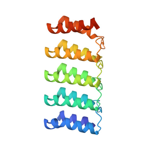
    In natural proteins, structured loops have central roles in molecular recognition, signal transduction and enzyme catalysis. However, because of the intrinsic flexibility and irregularity of loop regions, organizing multiple structured loops at protein functional sites has been very difficult to achieve by de novo protein design. Here we describe a solution to this problem that designs tandem repeat proteins with structured loops (9-14 residues) buttressed by extensive hydrogen bonding interactions. Experimental characterization shows that the designs are monodisperse, highly soluble, folded and thermally stable. Crystal structures are in close agreement with the design models, with the loops structured and buttressed as designed. We demonstrate the functionality afforded by loop buttressing by designing and characterizing binders for extended peptides in which the loops form one side of an extended binding pocket. The ability to design multiple structured loops should contribute generally to efforts to design new protein functions.


  • Organizational Affiliation
    • Department of Biochemistry, University of Washington, Seattle, WA, USA.

Macromolecule Content 

  • Total Structure Weight: 187.19 kDa 
  • Atom Count: 12,376 
  • Modeled Residue Count: 1,648 
  • Deposited Residue Count: 1,736 
  • Unique protein chains: 1

Macromolecules

Find similar proteins by:|  3D Structure
Entity ID: 1
MoleculeChains  Sequence LengthOrganismDetailsImage
RBL7_C2_3
A, B, C, D, E
A, B, C, D, E, F, G, H
217synthetic constructMutation(s): 0 

Small Molecules

Ligands 1 Unique
IDChains Name / Formula / InChI Key2D Diagram3D Interactions
MLI

Query on MLI


Download:Ideal Coordinates CCD File
I [auth B]MALONATE ION
C3 H2 O4
OFOBLEOULBTSOW-UHFFFAOYSA-L

Experimental Data & Validation

Experimental Data

  • Method: X-RAY DIFFRACTION
  • Resolution: 2.99 Å
  • R-Value Free:  0.259 (Depositor), 0.259 (DCC) 
  • R-Value Work:  0.210 (Depositor), 0.210 (DCC) 
  • R-Value Observed: 0.212 (Depositor) 
Space Group: P 21 21 21
Unit Cell:
Length ( Å )Angle ( ˚ )
a = 111.961α = 90
b = 117.644β = 90
c = 142.056γ = 90
Software Package:
Software NamePurpose
PHENIXrefinement
XDSdata reduction
XDSdata scaling
PHASERphasing

Structure Validation

View Full Validation Report


Entry History 

& Funding Information

Deposition Data


Funding OrganizationLocationGrant Number
Howard Hughes Medical Institute (HHMI)United States--

Revision History  (Full details and data files)

  • Version 1.0: 2024-04-17
    Type: Initial release
  • Version 1.1: 2024-06-26
    Changes: Database references
  • Version 1.2: 2024-08-07
    Changes: Database references