8FND

Structure of E138K HIV-1 intasome with Dolutegravir bound


Experimental Data Snapshot

  • Method: ELECTRON MICROSCOPY
  • Resolution: 3.00 Å
  • Aggregation State: PARTICLE 
  • Reconstruction Method: SINGLE PARTICLE 

wwPDB Validation   3D Report Full Report


This is version 1.1 of the entry. See complete history


Literature

Mechanisms of HIV-1 integrase resistance to dolutegravir and potent inhibition of drug-resistant variants.

Li, M.Oliveira Passos, D.Shan, Z.Smith, S.J.Sun, Q.Biswas, A.Choudhuri, I.Strutzenberg, T.S.Haldane, A.Deng, N.Li, Z.Zhao, X.Z.Briganti, L.Kvaratskhelia, M.Burke Jr., T.R.Levy, R.M.Hughes, S.H.Craigie, R.Lyumkis, D.

(2023) Sci Adv 9: eadg5953-eadg5953

  • DOI: https://doi.org/10.1126/sciadv.adg5953
  • Primary Citation of Related Structures:  
    8FN7, 8FND, 8FNG, 8FNH, 8FNJ, 8FNL, 8FNM, 8FNN, 8FNO, 8FNP, 8FNQ

  • PubMed Abstract: 

    HIV-1 infection depends on the integration of viral DNA into host chromatin. Integration is mediated by the viral enzyme integrase and is blocked by integrase strand transfer inhibitors (INSTIs), first-line antiretroviral therapeutics widely used in the clinic. Resistance to even the best INSTIs is a problem, and the mechanisms of resistance are poorly understood. Here, we analyze combinations of the mutations E138K, G140A/S, and Q148H/K/R, which confer resistance to INSTIs. The investigational drug 4d more effectively inhibited the mutants compared with the approved drug Dolutegravir (DTG). We present 11 new cryo-EM structures of drug-resistant HIV-1 intasomes bound to DTG or 4d, with better than 3-Å resolution. These structures, complemented with free energy simulations, virology, and enzymology, explain the mechanisms of DTG resistance involving E138K + G140A/S + Q148H/K/R and show why 4d maintains potency better than DTG. These data establish a foundation for further development of INSTIs that potently inhibit resistant forms in integrase.


  • Organizational Affiliation

    National Institute of Diabetes and Digestive Diseases, National Institutes of Health, Bethesda, MD, 20892, USA.


Macromolecules

Find similar proteins by:  (by identity cutoff)  |  3D Structure
Entity ID: 1
MoleculeChains Sequence LengthOrganismDetailsImage
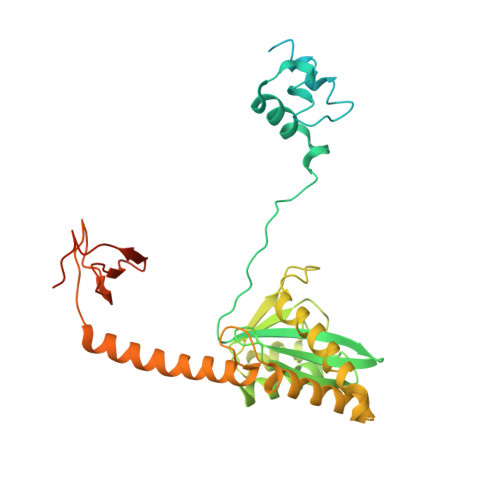
Lamina-associated polypeptide 2, isoform alpha,Integrase chimera
A, B, C, D, G
A, B, C, D, G, H, I, J
364Homo sapiensHuman immunodeficiency virus 1
This entity is chimeric
Mutation(s): 1 
Gene Names: TMPOLAP2gag-pol
EC: 2.7.7 (PDB Primary Data), 3.1 (PDB Primary Data)
UniProt & NIH Common Fund Data Resources
Find proteins for P42166 (Homo sapiens)
Explore P42166 
Go to UniProtKB:  P42166
PHAROS:  P42166
GTEx:  ENSG00000120802 
Find proteins for P12497 (Human immunodeficiency virus type 1 group M subtype B (isolate NY5))
Explore P12497 
Go to UniProtKB:  P12497
Entity Groups  
Sequence Clusters30% Identity50% Identity70% Identity90% Identity95% Identity100% Identity
UniProt GroupsP42166P12497
Sequence Annotations
Expand
  • Reference Sequence
Find similar nucleic acids by:  (by identity cutoff)  |  3D Structure
Entity ID: 2
MoleculeChains LengthOrganismImage
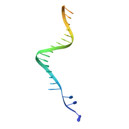
DNA (27-MER)
E, K
27Human immunodeficiency virus 1
Sequence Annotations
Expand
  • Reference Sequence

Find similar nucleic acids by:  Sequence   |   3D Structure  

Entity ID: 3
MoleculeChains LengthOrganismImage
DNA (25-MER)
F, L
25Human immunodeficiency virus 1
Sequence Annotations
Expand
  • Reference Sequence
Small Molecules
Ligands 3 Unique
IDChains Name / Formula / InChI Key2D Diagram3D Interactions
DLU (Subject of Investigation/LOI)
Query on DLU

Download Ideal Coordinates CCD File 
P [auth A],
T [auth G]
(4R,12aS)-N-(2,4-difluorobenzyl)-7-hydroxy-4-methyl-6,8-dioxo-3,4,6,8,12,12a-hexahydro-2H-pyrido[1',2':4,5]pyrazino[2,1-b][1,3]oxazine-9-carboxamide
C20 H19 F2 N3 O5
RHWKPHLQXYSBKR-BMIGLBTASA-N
ZN
Query on ZN

Download Ideal Coordinates CCD File 
O [auth A],
S [auth G]
ZINC ION
Zn
PTFCDOFLOPIGGS-UHFFFAOYSA-N
MG (Subject of Investigation/LOI)
Query on MG

Download Ideal Coordinates CCD File 
M [auth A],
N [auth A],
Q [auth G],
R [auth G]
MAGNESIUM ION
Mg
JLVVSXFLKOJNIY-UHFFFAOYSA-N
Experimental Data & Validation

Experimental Data

  • Method: ELECTRON MICROSCOPY
  • Resolution: 3.00 Å
  • Aggregation State: PARTICLE 
  • Reconstruction Method: SINGLE PARTICLE 
EM Software:
TaskSoftware PackageVersion
RECONSTRUCTIONcryoSPARC3
MODEL REFINEMENTPHENIX

Structure Validation

View Full Validation Report


Entry History & Funding Information

Deposition Data


Funding OrganizationLocationGrant Number
National Institutes of Health/National Institute Of Allergy and Infectious Diseases (NIH/NIAID)United StatesU01 AI136680
National Institutes of Health/National Institute Of Allergy and Infectious Diseases (NIH/NIAID)United StatesR01 AI146017
National Institutes of Health/National Institute Of Allergy and Infectious Diseases (NIH/NIAID)United StatesU54 AI170855
National Institutes of Health/National Institute of General Medical Sciences (NIH/NIGMS)United StatesR35 GM132090
National Institutes of Health/National Institute of General Medical Sciences (NIH/NIGMS)United StatesGM148049

Revision History  (Full details and data files)

  • Version 1.0: 2023-08-09
    Type: Initial release
  • Version 1.1: 2024-10-23
    Changes: Data collection, Structure summary