8FE6

Crystal structure of human O-GlcNAc transferase (OGT) in complex with an exosite-binding peptide and UDP-GlcNAc

  • Classification: TRANSFERASE
  • Organism(s): Homo sapiens
  • Expression System: Escherichia coli
  • Mutation(s): No 

  • Deposited: 2022-12-05 Released: 2023-08-09 
  • Deposition Author(s): Xie, J., Jiang, J.
  • Funding Organization(s): National Institutes of Health/National Institute of General Medical Sciences (NIH/NIGMS)

Experimental Data Snapshot

  • Method: X-RAY DIFFRACTION
  • Resolution: 3.06 Å
  • R-Value Free: 
    0.219 (Depositor), 0.220 (DCC) 
  • R-Value Work: 
    0.181 (Depositor), 0.180 (DCC) 
  • R-Value Observed: 
    0.182 (Depositor) 

Starting Model: experimental
View more details

wwPDB Validation   3D Report Full Report


Ligand Structure Quality Assessment 

Created with Raphaël 2.3.0Worse 01 BetterLigand structure goodness of fit to experimental dataBest fitted UD1Click on this verticalbar to view details

This is version 1.2 of the entry. See complete history


Literature

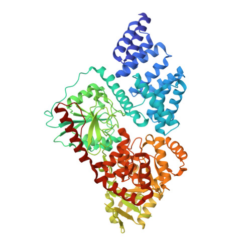
Motif-dependent binding on the intervening domain regulates O-GlcNAc transferase.

Blankenship, C.M.Xie, J.Benz, C.Wang, A.Ivarsson, Y.Jiang, J.

(2023) Nat Chem Biol 19: 1423-1431

  • DOI: https://doi.org/10.1038/s41589-023-01422-2
  • Primary Citation of Related Structures:  
    8FE6, 8FE7, 8FUF

  • PubMed Abstract: 

    The modification of intracellular proteins with O-linked β-N-acetylglucosamine (O-GlcNAc) moieties is a highly dynamic process that spatiotemporally regulates nearly every important cellular program. Despite its significance, little is known about the substrate recognition and regulation modes of O-GlcNAc transferase (OGT), the primary enzyme responsible for O-GlcNAc addition. In this study, we identified the intervening domain (Int-D), a poorly understood protein fold found only in metazoan OGTs, as a specific regulator of OGT protein-protein interactions and substrate modification. Using proteomic peptide phage display (ProP-PD) coupled with structural, biochemical and cellular characterizations, we discovered a strongly enriched peptide motif, employed by the Int-D to facilitate specific O-GlcNAcylation. We further show that disruption of Int-D binding dysregulates important cellular programs, including response to nutrient deprivation and glucose metabolism. These findings illustrate a mode of OGT substrate recognition and offer key insights into the biological roles of this unique domain.


  • Organizational Affiliation

    Pharmaceutical Sciences Division, School of Pharmacy, University of Wisconsin-Madison, Madison, WI, USA.


Macromolecules
Find similar proteins by:  (by identity cutoff)  |  3D Structure
Entity ID: 1
MoleculeChains Sequence LengthOrganismDetailsImage
UDP-N-acetylglucosamine--peptide N-acetylglucosaminyltransferase 110 kDa subunit
A, C, E, G
723Homo sapiensMutation(s): 0 
Gene Names: OGT
EC: 2.4.1.255
UniProt & NIH Common Fund Data Resources
Find proteins for O15294 (Homo sapiens)
Explore O15294 
Go to UniProtKB:  O15294
PHAROS:  O15294
GTEx:  ENSG00000147162 
Entity Groups  
Sequence Clusters30% Identity50% Identity70% Identity90% Identity95% Identity100% Identity
UniProt GroupO15294
Sequence Annotations
Expand
  • Reference Sequence

Find similar proteins by:  Sequence   |   3D Structure  

Entity ID: 2
MoleculeChains Sequence LengthOrganismDetailsImage
A motif peptide
B, D, F, H
12Homo sapiensMutation(s): 0 
Entity Groups  
Sequence Clusters30% Identity50% Identity70% Identity90% Identity95% Identity100% Identity
Sequence Annotations
Expand
  • Reference Sequence
Experimental Data & Validation

Experimental Data

  • Method: X-RAY DIFFRACTION
  • Resolution: 3.06 Å
  • R-Value Free:  0.219 (Depositor), 0.220 (DCC) 
  • R-Value Work:  0.181 (Depositor), 0.180 (DCC) 
  • R-Value Observed: 0.182 (Depositor) 
Space Group: H 3
Unit Cell:
Length ( Å )Angle ( ˚ )
a = 281.532α = 90
b = 281.532β = 90
c = 143.212γ = 120
Software Package:
Software NamePurpose
PHENIXrefinement
autoPROCdata processing
Cootmodel building
PHASERphasing
XDSdata reduction
Aimlessdata scaling

Structure Validation

View Full Validation Report


Ligand Structure Quality Assessment 

Created with Raphaël 2.3.0Worse 01 BetterLigand structure goodness of fit to experimental dataBest fitted UD1Click on this verticalbar to view details

Entry History & Funding Information

Deposition Data


Funding OrganizationLocationGrant Number
National Institutes of Health/National Institute of General Medical Sciences (NIH/NIGMS)United StatesGM121718

Revision History  (Full details and data files)

  • Version 1.0: 2023-08-09
    Type: Initial release
  • Version 1.1: 2023-11-01
    Changes: Data collection, Database references
  • Version 1.2: 2023-11-08
    Changes: Database references