8F99

Compound 10 bound to procaspase-6


Experimental Data Snapshot

  • Method: X-RAY DIFFRACTION
  • Resolution: 2.86 Å
  • R-Value Free: 0.246 
  • R-Value Work: 0.200 
  • R-Value Observed: 0.202 

wwPDB Validation   3D Report Full Report


Ligand Structure Quality Assessment 


This is version 1.1 of the entry. See complete history


Literature

Systematic Study of Heteroarene Stacking Using a Congeneric Set of Molecular Glues for Procaspase-6.

Togo, T.Tram, L.Denton, L.G.ElHilali-Pollard, X.Gu, J.Jiang, J.Liu, C.Zhao, Y.Zhao, Y.Zheng, Y.Zheng, Y.Yang, J.Fan, P.Arkin, M.R.Harma, H.Sun, D.Canan, S.S.Wheeler, S.E.Renslo, A.R.

(2023) J Med Chem 66: 9784-9796

  • DOI: https://doi.org/10.1021/acs.jmedchem.3c00590
  • Primary Citation of Related Structures:  
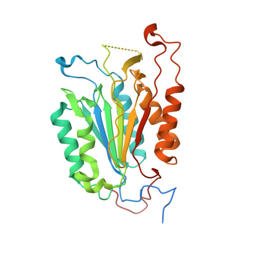
    8F78, 8F96, 8F97, 8F98, 8F99, 8F9A, 8F9B, 8F9C, 8F9D, 8FBV

  • PubMed Abstract: 

    Heteroaromatic stacking interactions are important in drug binding, supramolecular chemistry, and materials science, making protein-ligand model systems of these interactions of considerable interest. Here we studied 30 congeneric ligands that each present a distinct heteroarene for stacking between tyrosine residues at the dimer interface of procaspase-6. Complex X-ray crystal structures of 10 analogs showed that stacking geometries were well conserved, while high-accuracy computations showed that heteroarene stacking energy was well correlated with predicted overall ligand binding energies. Empirically determined K D values in this system thus provide a useful measure of heteroarene stacking with tyrosine. Stacking energies are discussed in the context of torsional strain, the number and positioning of heteroatoms, tautomeric state, and coaxial orientation of heteroarene in the stack. Overall, this study provides an extensive data set of empirical and high-level computed binding energies in a versatile new protein-ligand system amenable to studies of other intermolecular interactions.


  • Organizational Affiliation

    Department of Pharmaceutical Chemistry, University of California, 600 16th Street, San Francisco, California 94143, United States.


Macromolecules
Find similar proteins by:  (by identity cutoff)  |  3D Structure
Entity ID: 1
MoleculeChains Sequence LengthOrganismDetailsImage
Procaspase-6
A, B, C, D
299Homo sapiensMutation(s): 0 
Gene Names: CASP6MCH2
EC: 3.4.22.59
UniProt & NIH Common Fund Data Resources
Find proteins for P55212 (Homo sapiens)
Explore P55212 
Go to UniProtKB:  P55212
PHAROS:  P55212
GTEx:  ENSG00000138794 
Entity Groups  
Sequence Clusters30% Identity50% Identity70% Identity90% Identity95% Identity100% Identity
UniProt GroupP55212
Sequence Annotations
Expand
  • Reference Sequence
Small Molecules
Ligands 1 Unique
IDChains Name / Formula / InChI Key2D Diagram3D Interactions
XMR (Subject of Investigation/LOI)
Query on XMR

Download Ideal Coordinates CCD File 
E [auth A],
F [auth C]
5-fluoro-2-({[(3M)-3-(1,3-oxazol-4-yl)pyridin-2-yl]amino}methyl)phenol
C15 H12 F N3 O2
AIPASLMZJHFYBT-UHFFFAOYSA-N
Experimental Data & Validation

Experimental Data

  • Method: X-RAY DIFFRACTION
  • Resolution: 2.86 Å
  • R-Value Free: 0.246 
  • R-Value Work: 0.200 
  • R-Value Observed: 0.202 
  • Space Group: P 61
Unit Cell:
Length ( Å )Angle ( ˚ )
a = 101.481α = 90
b = 101.481β = 90
c = 321.539γ = 120
Software Package:
Software NamePurpose
Aimlessdata scaling
REFMACrefinement
PDB_EXTRACTdata extraction
XDSdata reduction
PHASERphasing

Structure Validation

View Full Validation Report


Ligand Structure Quality Assessment 


Entry History & Funding Information

Deposition Data


Funding OrganizationLocationGrant Number
Not funded--

Revision History  (Full details and data files)

  • Version 1.0: 2023-12-13
    Type: Initial release
  • Version 1.1: 2024-06-26
    Changes: Database references