8F6P | pdb_00008f6p

Rat Cardiac Sodium Channel with Ranolazine Bound


Experimental Data Snapshot

  • Method: ELECTRON MICROSCOPY
  • Resolution: 3.20 Å
  • Aggregation State: PARTICLE 
  • Reconstruction Method: SINGLE PARTICLE 

wwPDB Validation 3D Report Full Report

Validation slider image for 8F6P

This is version 1.2 of the entry. See complete history

Literature

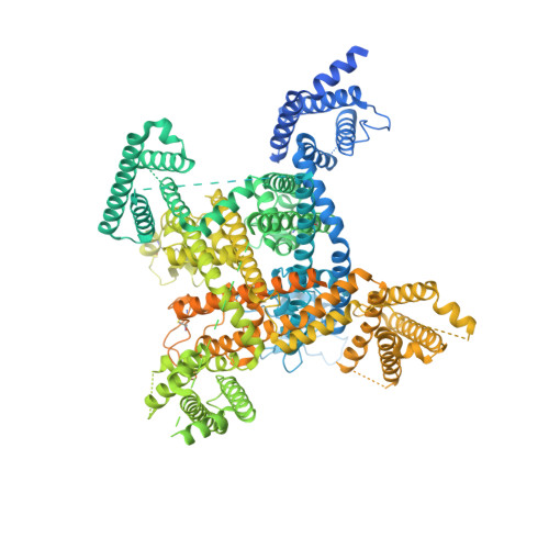
Structural basis for inhibition of the cardiac sodium channel by the atypical antiarrhythmic drug ranolazine.

Lenaeus, M.Gamal El-Din, T.M.Tonggu, L.Zheng, N.Catterall, W.A.

(2023) Nat Cardiovasc Res 2: 587-594

Macromolecule Content 

  • Total Structure Weight: 223.1 kDa 
  • Atom Count: 9,278 
  • Modeled Residue Count: 1,091 
  • Deposited Residue Count: 1,838 
  • Unique protein chains: 1

Macromolecules

Find similar proteins by:|  3D Structure
Entity ID: 1
MoleculeChains  Sequence LengthOrganismDetailsImage
Sodium channel protein type 5 subunit alpha,Green fluorescent protein1,838Rattus norvegicusAequorea victoriaMutation(s): 0 
Gene Names: Scn5aGFP
Membrane Entity: Yes 
UniProt
Find proteins for P15389 (Rattus norvegicus)
Explore P15389 
Go to UniProtKB:  P15389
Find proteins for P42212 (Aequorea victoria)
Explore P42212 
Go to UniProtKB:  P42212
Entity Groups
Sequence Clusters30% Identity50% Identity70% Identity90% Identity95% Identity100% Identity
UniProt GroupsP42212P15389
Glycosylation
Glycosylation Sites: 3Go to GlyGen: P15389-1
Sequence Annotations
Expand
Reference Sequence

Oligosaccharides

Help  
Entity ID: 2
MoleculeChains Length2D Diagram GlycosylationD Interactions
2-acetamido-2-deoxy-beta-D-glucopyranose-(1-4)-2-acetamido-2-deoxy-beta-D-glucopyranose
B, C
2N-Glycosylation
Glycosylation Resources
GlyTouCan: G42666HT
GlyCosmos: G42666HT
GlyGen: G42666HT

Small Molecules

Ligands 5 Unique
IDChains Name / Formula / InChI Key2D Diagram3D Interactions
6OU

Query on 6OU


Download:Ideal Coordinates CCD File
O [auth A]
P [auth A]
Q [auth A]
R [auth A]
S [auth A]
O [auth A],
P [auth A],
Q [auth A],
R [auth A],
S [auth A],
T [auth A]
[(2~{R})-1-[2-azanylethoxy(oxidanyl)phosphoryl]oxy-3-hexadecanoyloxy-propan-2-yl] (~{Z})-octadec-9-enoate
C39 H76 N O8 P
FHQVHHIBKUMWTI-OTMQOFQLSA-N
Y01

Query on Y01


Download:Ideal Coordinates CCD File
AA [auth A]
BA [auth A]
CA [auth A]
DA [auth A]
EA [auth A]
AA [auth A],
BA [auth A],
CA [auth A],
DA [auth A],
EA [auth A],
FA [auth A],
GA [auth A],
U [auth A],
V [auth A],
W [auth A],
X [auth A],
Y [auth A],
Z [auth A]
CHOLESTEROL HEMISUCCINATE
C31 H50 O4
WLNARFZDISHUGS-MIXBDBMTSA-N
XHO
(Subject of Investigation/LOI)

Query on XHO


Download:Ideal Coordinates CCD File
N [auth A](R)-ranolazine
C24 H33 N3 O4
XKLMZUWKNUAPSZ-HXUWFJFHSA-N
NAG

Query on NAG


Download:Ideal Coordinates CCD File
D [auth A]
E [auth A]
F [auth A]
H [auth A]
K [auth A]
D [auth A],
E [auth A],
F [auth A],
H [auth A],
K [auth A],
L [auth A],
M [auth A]
2-acetamido-2-deoxy-beta-D-glucopyranose
C8 H15 N O6
OVRNDRQMDRJTHS-FMDGEEDCSA-N
BMA

Query on BMA


Download:Ideal Coordinates CCD File
G [auth A],
I [auth A],
J [auth A]
beta-D-mannopyranose
C6 H12 O6
WQZGKKKJIJFFOK-RWOPYEJCSA-N

Experimental Data & Validation

Experimental Data

  • Method: ELECTRON MICROSCOPY
  • Resolution: 3.20 Å
  • Aggregation State: PARTICLE 
  • Reconstruction Method: SINGLE PARTICLE 
EM Software:
TaskSoftware PackageVersion
MODEL REFINEMENTPHENIX

Structure Validation

View Full Validation Report


Entry History 

& Funding Information

Deposition Data


Funding OrganizationLocationGrant Number
National Institutes of Health/National Heart, Lung, and Blood Institute (NIH/NHLBI)United StatesK08 HL145630-05

Revision History  (Full details and data files)

  • Version 1.0: 2023-04-12
    Type: Initial release
  • Version 1.1: 2024-10-16
    Changes: Data collection, Database references, Structure summary
  • Version 1.2: 2024-11-13
    Changes: Data collection