8F68 | pdb_00008f68

E. coli cytochrome bo3 ubiquinol oxidase monomer


Experimental Data Snapshot

  • Method: ELECTRON MICROSCOPY
  • Resolution: 3.15 Å
  • Aggregation State: PARTICLE 
  • Reconstruction Method: SINGLE PARTICLE 

wwPDB Validation 3D Report Full Report

Validation slider image for 8F68

This is version 1.5 of the entry. See complete history

Literature

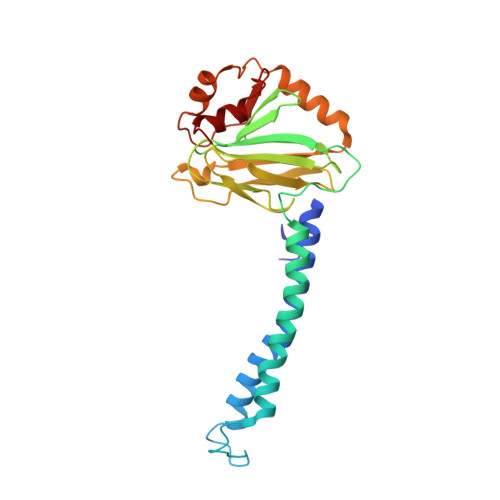
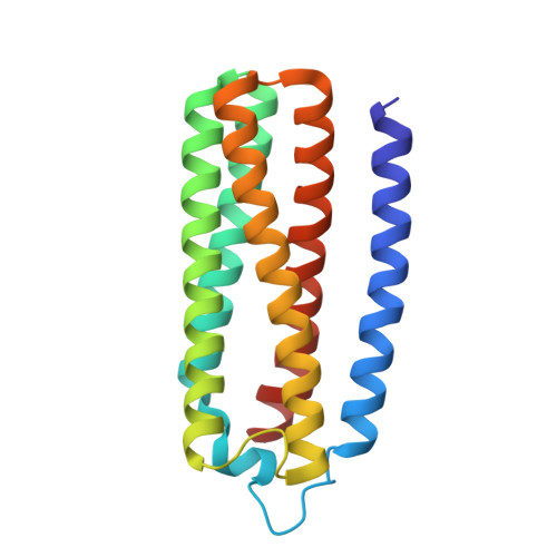
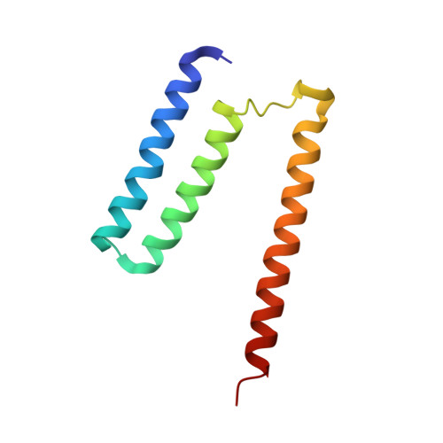
Monomer and dimer structures of cytochrome bo 3 ubiquinol oxidase from Escherichia coli.

Guo, Y.Karimullina, E.Emde, T.Otwinowski, Z.Borek, D.Savchenko, A.

(2023) Protein Sci 32: e4616-e4616

  • DOI: https://doi.org/10.1002/pro.4616
  • Primary Citation Related Structures: 
    8F68, 8F6C

  • PubMed Abstract: 

    The Escherichia coli cytochrome bo 3 ubiquinol oxidase is a four-subunit heme-copper oxidase that serves as a proton pump in the E. coli aerobic respiratory chain. Despite many mechanistic studies, it is unclear whether this ubiquinol oxidase functions as a monomer, or as a dimer in a manner similar to its eukaryotic counterparts-the mitochondrial electron transport complexes. In this study, we determined the monomeric and dimeric structures of the E. coli cytochrome bo 3 ubiquinol oxidase reconstituted in amphipol by cryogenic electron microscopy single particle reconstruction (cryo-EM SPR) to a resolution of 3.15 and 3.46 Å, respectively. We have discovered that the protein can form a dimer with C2 symmetry, with the dimerization interface maintained by interactions between the subunit II of one monomer and the subunit IV of the other monomer. Moreover, the dimerization does not induce significant structural changes in the monomers, except the movement of a loop in subunit IV (residues 67-74).


  • Organizational Affiliation
    • Department of Biophysics, The University of Texas Southwestern Medical Center, Dallas, Texas, USA.

Macromolecule Content 

  • Total Structure Weight: 137.62 kDa 
  • Atom Count: 9,666 
  • Modeled Residue Count: 1,198 
  • Deposited Residue Count: 1,198 
  • Unique protein chains: 4

Macromolecules

Find similar proteins by:|  3D Structure
Entity ID: 1
MoleculeChains  Sequence LengthOrganismDetailsImage
Cytochrome bo(3) ubiquinol oxidase subunit 1658Escherichia coliMutation(s): 0 
Gene Names: cyoBb0431JW0421
EC: 7.1.1.3
Membrane Entity: Yes 
UniProt
Find proteins for P0ABI8 (Escherichia coli (strain K12))
Explore P0ABI8 
Go to UniProtKB:  P0ABI8
Entity Groups
Sequence Clusters30% Identity50% Identity70% Identity90% Identity95% Identity100% Identity
UniProt GroupP0ABI8
Sequence Annotations
Expand
Reference Sequence
Find similar proteins by:|  3D Structure
Entity ID: 2
MoleculeChains  Sequence LengthOrganismDetailsImage
Cytochrome bo(3) ubiquinol oxidase subunit 2260Escherichia coliMutation(s): 0 
Gene Names: cyoAb0432JW0422
Membrane Entity: Yes 
UniProt
Find proteins for P0ABJ1 (Escherichia coli (strain K12))
Explore P0ABJ1 
Go to UniProtKB:  P0ABJ1
Entity Groups
Sequence Clusters30% Identity50% Identity70% Identity90% Identity95% Identity100% Identity
UniProt GroupP0ABJ1
Sequence Annotations
Expand
Reference Sequence
Find similar proteins by:|  3D Structure
Entity ID: 3
MoleculeChains  Sequence LengthOrganismDetailsImage
Cytochrome bo(3) ubiquinol oxidase subunit 3184Escherichia coliMutation(s): 0 
Gene Names: cyoCb0430JW0420
Membrane Entity: Yes 
UniProt
Find proteins for P0ABJ3 (Escherichia coli (strain K12))
Explore P0ABJ3 
Go to UniProtKB:  P0ABJ3
Entity Groups
Sequence Clusters30% Identity50% Identity70% Identity90% Identity95% Identity100% Identity
UniProt GroupP0ABJ3
Sequence Annotations
Expand
Reference Sequence
Find similar proteins by:|  3D Structure
Entity ID: 4
MoleculeChains  Sequence LengthOrganismDetailsImage
Cytochrome bo(3) ubiquinol oxidase subunit 496Escherichia coliMutation(s): 0 
Gene Names: cyoDb0429JW0419
Membrane Entity: Yes 
UniProt
Find proteins for P0ABJ6 (Escherichia coli (strain K12))
Explore P0ABJ6 
Go to UniProtKB:  P0ABJ6
Entity Groups
Sequence Clusters30% Identity50% Identity70% Identity90% Identity95% Identity100% Identity
UniProt GroupP0ABJ6
Sequence Annotations
Expand
Reference Sequence

Small Molecules

Ligands 4 Unique
IDChains Name / Formula / InChI Key2D Diagram3D Interactions
HEO
(Subject of Investigation/LOI)

Query on HEO


Download:Ideal Coordinates CCD File
F [auth A]HEME O
C49 H58 Fe N4 O5
FRKORVCRVCLRBA-BZKSIRDQSA-L
3PE

Query on 3PE


Download:Ideal Coordinates CCD File
H [auth A],
I [auth A],
J [auth C]
1,2-Distearoyl-sn-glycerophosphoethanolamine
C41 H82 N O8 P
LVNGJLRDBYCPGB-LDLOPFEMSA-N
HEM
(Subject of Investigation/LOI)

Query on HEM


Download:Ideal Coordinates CCD File
E [auth A]PROTOPORPHYRIN IX CONTAINING FE
C34 H32 Fe N4 O4
KABFMIBPWCXCRK-RGGAHWMASA-L
CU

Query on CU


Download:Ideal Coordinates CCD File
G [auth A]COPPER (II) ION
Cu
JPVYNHNXODAKFH-UHFFFAOYSA-N

Experimental Data & Validation

Experimental Data

  • Method: ELECTRON MICROSCOPY
  • Resolution: 3.15 Å
  • Aggregation State: PARTICLE 
  • Reconstruction Method: SINGLE PARTICLE 

Structure Validation

View Full Validation Report


Entry History 

& Funding Information

Deposition Data


Funding OrganizationLocationGrant Number
National Institutes of Health/National Institute Of Allergy and Infectious Diseases (NIH/NIAID)United States--

Revision History  (Full details and data files)

  • Version 1.0: 2022-11-30
    Type: Initial release
  • Version 1.1: 2023-03-22
    Changes: Database references
  • Version 1.2: 2023-04-12
    Changes: Database references
  • Version 1.3: 2023-04-19
    Changes: Database references
  • Version 1.4: 2024-05-22
    Changes: Data collection
  • Version 1.5: 2025-09-17
    Changes: Data collection, Database references, Other, Structure summary