


Structure of scaffolidng protein D13 of Vaccinia Virus in complex with fragments inhibiting A17 binding.
Subedi, B.P., Garriga, D., Coulibaly, F.To be published.
Experimental Data Snapshot
Starting Model: experimental
View more details
Entity ID: 1 | |||||
|---|---|---|---|---|---|
| Molecule | Chains | Sequence Length | Organism | Details | Image |
| Scaffold protein D13 | 576 | Orthopoxvirus vaccinia | Mutation(s): 0 Gene Names: VACWR118, D13L | 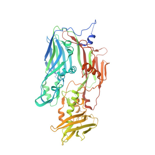 | |
UniProt | |||||
Entity Groups | |||||
| Sequence Clusters | 30% Identity50% Identity70% Identity90% Identity95% Identity100% Identity | ||||
| UniProt Group | P68440 | ||||
Sequence AnnotationsExpand | |||||
Reference Sequence | |||||
| Ligands 2 Unique | |||||
|---|---|---|---|---|---|
| ID | Chains | Name / Formula / InChI Key | 2D Diagram | 3D Interactions | |
| QF6 (Subject of Investigation/LOI) Download:Ideal Coordinates CCD File | Y [auth B] | 8-methoxyquinolin-4-amine C10 H10 N2 O QMBPJEIUEYDRGP-UHFFFAOYSA-N |  | ||
| FMT Download:Ideal Coordinates CCD File | AA [auth C] BA [auth C] CA [auth C] D [auth A] DA [auth C] | FORMIC ACID C H2 O2 BDAGIHXWWSANSR-UHFFFAOYSA-N |  | ||
| Length ( Å ) | Angle ( ˚ ) |
|---|---|
| a = 191.078 | α = 90 |
| b = 191.078 | β = 90 |
| c = 254.717 | γ = 120 |
| Software Name | Purpose |
|---|---|
| PHENIX | refinement |
| BUSTER | refinement |
| XDS | data reduction |
| Aimless | data scaling |
| PHASER | phasing |
| Funding Organization | Location | Grant Number |
|---|---|---|
| National Health and Medical Research Council (NHMRC, Australia) | Australia | APP1051907 |


