8EYW | pdb_00008eyw

Beetroot dimer bound to ThT


Experimental Data Snapshot

  • Method: X-RAY DIFFRACTION
  • Resolution: 2.10 Å
  • R-Value Free: 
    0.220 (Depositor), 0.220 (DCC) 
  • R-Value Work: 
    0.180 (Depositor), 0.180 (DCC) 
  • R-Value Observed: 
    0.183 (Depositor) 

Starting Model: experimental
View more details

wwPDB Validation 3D Report Full Report

Validation slider image for 8EYW

Ligand Structure Quality Assessment 


This is version 1.2 of the entry. See complete history

Literature

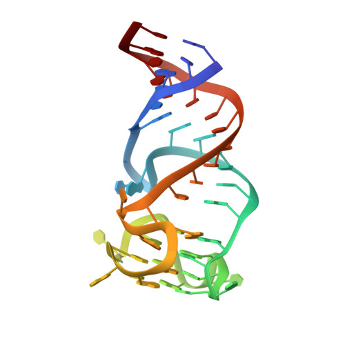
Co-crystal structures of the fluorogenic aptamer Beetroot show that close homology may not predict similar RNA architecture.

Passalacqua, L.F.M.Starich, M.R.Link, K.A.Wu, J.Knutson, J.R.Tjandra, N.Jaffrey, S.R.Ferre-D'Amare, A.R.

(2023) Nat Commun 14: 2969-2969

  • DOI: https://doi.org/10.1038/s41467-023-38683-3
  • Primary Citation Related Structures: 
    8EYU, 8EYV, 8EYW, 8F0N

  • PubMed Abstract: 

    Beetroot is a homodimeric in vitro selected RNA that binds and activates DFAME, a conditional fluorophore derived from GFP. It is 70% sequence-identical to the previously characterized homodimeric aptamer Corn, which binds one molecule of its cognate fluorophore DFHO at its interprotomer interface. We have now determined the Beetroot-DFAME co-crystal structure at 1.95 Å resolution, discovering that this RNA homodimer binds two molecules of the fluorophore, at sites separated by ~30 Å. In addition to this overall architectural difference, the local structures of the non-canonical, complex quadruplex cores of Beetroot and Corn are distinctly different, underscoring how subtle RNA sequence differences can give rise to unexpected structural divergence. Through structure-guided engineering, we generated a variant that has a 12-fold fluorescence activation selectivity switch toward DFHO. Beetroot and this variant form heterodimers and constitute the starting point for engineered tags whose through-space inter-fluorophore interaction could be used to monitor RNA dimerization.


  • Organizational Affiliation
    • Biochemistry and Biophysics Center, National Heart, Lung, and Blood Institute, National Institutes of Health, Bethesda, MD, USA.

Macromolecule Content 

  • Total Structure Weight: 32.81 kDa 
  • Atom Count: 2,221 
  • Modeled Residue Count: 94 
  • Deposited Residue Count: 98 
  • Unique nucleic acid chains: 1

Macromolecules

Find similar nucleic acids by:  (by identity cutoff) 
Entity ID: 1
MoleculeChains LengthOrganismImage
RNA (49-MER)A [auth B],
B [auth A]
49synthetic construct
Sequence Annotations
Expand
Reference Sequence

Experimental Data & Validation

Experimental Data

  • Method: X-RAY DIFFRACTION
  • Resolution: 2.10 Å
  • R-Value Free:  0.220 (Depositor), 0.220 (DCC) 
  • R-Value Work:  0.180 (Depositor), 0.180 (DCC) 
  • R-Value Observed: 0.183 (Depositor) 
Space Group: P 1 21 1
Unit Cell:
Length ( Å )Angle ( ˚ )
a = 29.687α = 90
b = 50.566β = 93.184
c = 94.74γ = 90
Software Package:
Software NamePurpose
PHENIXrefinement
Cootmodel building
xia2data reduction
DIALSdata scaling

Structure Validation

View Full Validation Report


Ligand Structure Quality Assessment 


Entry History 

& Funding Information

Deposition Data


Funding OrganizationLocationGrant Number
National Institutes of Health/National Heart, Lung, and Blood Institute (NIH/NHLBI)United States--

Revision History  (Full details and data files)

  • Version 1.0: 2023-05-31
    Type: Initial release
  • Version 1.1: 2023-09-20
    Changes: Data collection, Database references
  • Version 1.2: 2023-10-25
    Changes: Refinement description