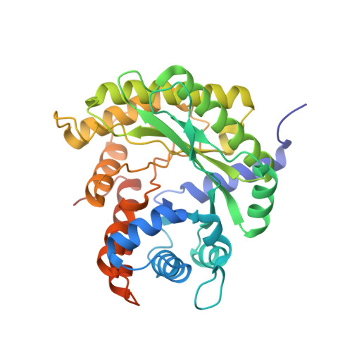
Addressing compressive deformation of proteins embedded in crystalline ice.
Shi, H., Wu, C., Zhang, X.(2023) Structure 31: 213
- PubMed: 36586403
- DOI: https://doi.org/10.1016/j.str.2022.12.001
- Primary Citation of Related Structures:
8BQN, 8EW0, 8EW2, 8F49, 8F7Y, 8HHS, 8HI2 - PubMed Abstract:
For cryoelectron microscopy (cryo-EM), high cooling rates have been required for preparation of protein samples to vitrify the surrounding water and avoid formation of damaging crystalline ice. Whether and how crystalline ice affects single-particle cryo-EM is still unclear. Here, single-particle cryo-EM was used to analyze three-dimensional structures of various proteins and viruses embedded in crystalline ice formed at various cooling rates. Low cooling rates led to shrinkage deformation and density distortions on samples having loose structures. Higher cooling rates reduced deformations. Deformation-free proteins in crystalline ice were obtained by modifying the freezing conditions, and reconstructions from these samples revealed a marked improvement over vitreous ice. This procedure also increased the efficiency of cryo-EM structure determinations and was essential for high-resolution reconstructions.
Organizational Affiliation:
National Laboratory of Biomacromolecules, CAS Center for Excellence in Biomacromolecules, Institute of Biophysics, Chinese Academy of Sciences (CAS), Beijing 100101, P.R. China; University of Chinese Academy of Sciences, Beijing 100049, P.R. China.


