8ER0 | pdb_00008er0

X-ray crystal structure of Tet(X6) bound to anhydrotetracycline


Experimental Data Snapshot

  • Method: X-RAY DIFFRACTION
  • Resolution: 2.20 Å
  • R-Value Free: 
    0.231 (Depositor), 0.231 (DCC) 
  • R-Value Work: 
    0.193 (Depositor), 0.194 (DCC) 
  • R-Value Observed: 
    0.194 (Depositor) 

Starting Model: experimental
View more details

wwPDB Validation 3D Report Full Report

Validation slider image for 8ER0

Ligand Structure Quality Assessment 


This is version 1.1 of the entry. See complete history

Literature

Structure of anhydrotetracycline-bound Tet(X6) reveals the mechanism for inhibition of type 1 tetracycline destructases.

Kumar, H.Williford, E.E.Blake, K.S.Virgin-Downey, B.Dantas, G.Wencewicz, T.A.Tolia, N.H.

(2023) Commun Biol 6: 423-423

  • DOI: https://doi.org/10.1038/s42003-023-04792-4
  • Primary Citation Related Structures: 
    8ER0, 8ER1

  • PubMed Abstract: 

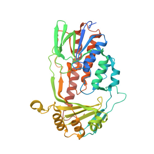
    Inactivation of tetracycline antibiotics by tetracycline destructases (TDases) remains a clinical and agricultural threat. TDases can be classified as type 1 Tet(X)-like TDases and type 2 soil-derived TDases. Type 1 TDases are widely identified in clinical pathogens. A combination therapy of tetracycline and a TDase inhibitor is much needed to rescue the clinical efficacy of tetracyclines. Anhydrotetracycline is a pan-TDase inhibitor that inhibits both type 1 and type 2 TDases. Here, we present structural, biochemical, and phenotypic evidence that anhydrotetracycline binds in a substrate-like orientation and competitively inhibits the type 1 TDase Tet(X6) to rescue tetracycline antibiotic activity as a sacrificial substrate. Anhydrotetracycline interacting residues of Tet(X6) are conserved within type 1 TDases, indicating a conserved binding mode and mechanism of inhibition. This mode of binding and inhibition is distinct from anhydrotetracycline's inhibition of type 2 TDases. This study forms the framework for development of next-generation therapies to counteract enzymatic tetracycline resistance.


  • Organizational Affiliation
    • Host-pathogen interaction and structural vaccinology section (HPISV), National Institute of Allergy and Infectious Diseases (NIAID), National Institutes of Health (NIH), Bethesda, MD, USA.

Macromolecule Content 

  • Total Structure Weight: 93.72 kDa 
  • Atom Count: 6,308 
  • Modeled Residue Count: 739 
  • Deposited Residue Count: 806 
  • Unique protein chains: 1

Macromolecules

Find similar proteins by:|  3D Structure
Entity ID: 1
MoleculeChains  Sequence LengthOrganismDetailsImage
Flavin-dependent monooxygenase
A, B
403Chryseobacterium oncorhynchiMutation(s): 0 
Gene Names: tet(X)
EC: 1.14.13
UniProt
Find proteins for A0A316WTJ0 (Chryseobacterium oncorhynchi)
Explore A0A316WTJ0 
Go to UniProtKB:  A0A316WTJ0
Entity Groups
Sequence Clusters30% Identity50% Identity70% Identity90% Identity95% Identity100% Identity
UniProt GroupA0A316WTJ0
Sequence Annotations
Expand
Reference Sequence

Experimental Data & Validation

Experimental Data

  • Method: X-RAY DIFFRACTION
  • Resolution: 2.20 Å
  • R-Value Free:  0.231 (Depositor), 0.231 (DCC) 
  • R-Value Work:  0.193 (Depositor), 0.194 (DCC) 
  • R-Value Observed: 0.194 (Depositor) 
Space Group: P 1 21 1
Unit Cell:
Length ( Å )Angle ( ˚ )
a = 87.086α = 90
b = 52.212β = 95.44
c = 94.871γ = 90
Software Package:
Software NamePurpose
PHENIXrefinement
XDSdata reduction
Aimlessdata scaling
PHASERphasing

Structure Validation

View Full Validation Report


Ligand Structure Quality Assessment 


Entry History 

& Funding Information

Deposition Data


Funding OrganizationLocationGrant Number
National Institutes of Health/National Institute Of Allergy and Infectious Diseases (NIH/NIAID)United States2U01AI123394
National Institutes of Health/National Institute Of Allergy and Infectious Diseases (NIH/NIAID)United StatesIntramural Research Program

Revision History  (Full details and data files)

  • Version 1.0: 2023-04-26
    Type: Initial release
  • Version 1.1: 2023-10-25
    Changes: Data collection, Refinement description