8ELI

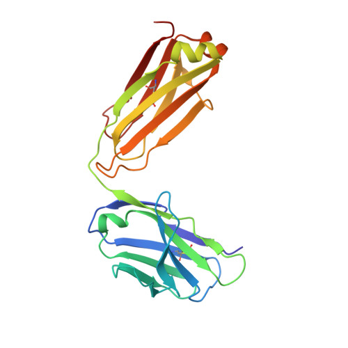
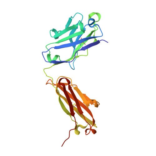
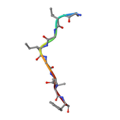
Broadly neutralizing antibody VRC34-combo.1 in complex with HIV fusion peptide (residue 512-519)


Experimental Data Snapshot

  • Method: X-RAY DIFFRACTION
  • Resolution: 1.49 Å
  • R-Value Free: 0.178 
  • R-Value Work: 0.162 
  • R-Value Observed: 0.163 

wwPDB Validation   3D Report Full Report


This is version 1.1 of the entry. See complete history


Literature

Antibody-directed evolution reveals a mechanism for enhanced neutralization at the HIV-1 fusion peptide site.

Banach, B.B.Pletnev, S.Olia, A.S.Xu, K.Zhang, B.Rawi, R.Bylund, T.Doria-Rose, N.A.Nguyen, T.D.Fahad, A.S.Lee, M.Lin, B.C.Liu, T.Louder, M.K.Madan, B.McKee, K.O'Dell, S.Sastry, M.Schon, A.Bui, N.Shen, C.H.Wolfe, J.R.Chuang, G.Y.Mascola, J.R.Kwong, P.D.DeKosky, B.J.

(2023) Nat Commun 14: 7593-7593

  • DOI: https://doi.org/10.1038/s41467-023-42098-5
  • Primary Citation of Related Structures:  
    8ELI, 8EUU, 8EUV, 8EUW, 8F7Z

  • PubMed Abstract: 

    The HIV-1 fusion peptide (FP) represents a promising vaccine target, but global FP sequence diversity among circulating strains has limited anti-FP antibodies to ~60% neutralization breadth. Here we evolve the FP-targeting antibody VRC34.01 in vitro to enhance FP-neutralization using site saturation mutagenesis and yeast display. Successive rounds of directed evolution by iterative selection of antibodies for binding to resistant HIV-1 strains establish a variant, VRC34.01_mm28, as a best-in-class antibody with 10-fold enhanced potency compared to the template antibody and ~80% breadth on a cross-clade 208-strain neutralization panel. Structural analyses demonstrate that the improved paratope expands the FP binding groove to accommodate diverse FP sequences of different lengths while also recognizing the HIV-1 Env backbone. These data reveal critical antibody features for enhanced neutralization breadth and potency against the FP site of vulnerability and accelerate clinical development of broad HIV-1 FP-targeting vaccines and therapeutics.


  • Organizational Affiliation

    Bioengineering Graduate Program, The University of Kansas, Lawrence, KS, 66045, USA.


Macromolecules
Find similar proteins by:  (by identity cutoff)  |  3D Structure
Entity ID: 1
MoleculeChains Sequence LengthOrganismDetailsImage
VRC34-combo.1 Fab Light chainA [auth L]213Homo sapiensMutation(s): 0 
Entity Groups  
Sequence Clusters30% Identity50% Identity70% Identity90% Identity95% Identity100% Identity
Sequence Annotations
Expand
  • Reference Sequence
Find similar proteins by:  (by identity cutoff)  |  3D Structure
Entity ID: 2
MoleculeChains Sequence LengthOrganismDetailsImage
VRC34-combo.1 Fab Heavy chainB [auth H]226Homo sapiensMutation(s): 0 
Entity Groups  
Sequence Clusters30% Identity50% Identity70% Identity90% Identity95% Identity100% Identity
Sequence Annotations
Expand
  • Reference Sequence

Find similar proteins by:  Sequence   |   3D Structure  

Entity ID: 3
MoleculeChains Sequence LengthOrganismDetailsImage
Fusion peptideC [auth A]8HIV-1 06TG.HT008Mutation(s): 0 
UniProt
Find proteins for P04624 (Human immunodeficiency virus type 1 group M subtype B (isolate HXB3))
Explore P04624 
Go to UniProtKB:  P04624
Entity Groups  
UniProt GroupP04624
Sequence Annotations
Expand
  • Reference Sequence
Experimental Data & Validation

Experimental Data

  • Method: X-RAY DIFFRACTION
  • Resolution: 1.49 Å
  • R-Value Free: 0.178 
  • R-Value Work: 0.162 
  • R-Value Observed: 0.163 
  • Space Group: P 21 21 21
Unit Cell:
Length ( Å )Angle ( ˚ )
a = 38.323α = 90
b = 86.152β = 90
c = 128.824γ = 90
Software Package:
Software NamePurpose
HKL-2000data scaling
PHENIXrefinement
PDB_EXTRACTdata extraction
HKL-2000data reduction
PHASERphasing

Structure Validation

View Full Validation Report


Entry History & Funding Information

Deposition Data


Funding OrganizationLocationGrant Number
Not funded--

Revision History  (Full details and data files)

  • Version 1.0: 2023-09-27
    Type: Initial release
  • Version 1.1: 2024-02-14
    Changes: Database references