8EFI | pdb_00008efi

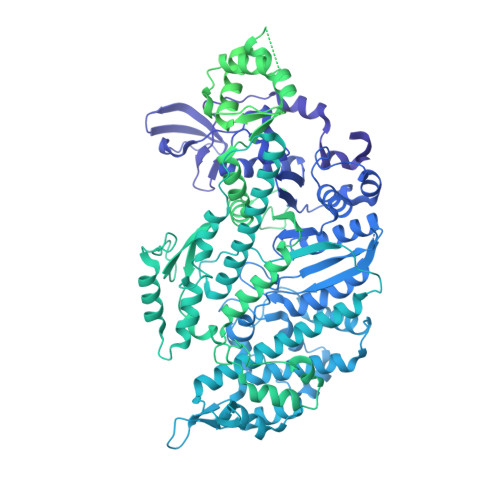
Helical reconstruction of the human cardiac actin-tropomyosin-myosin complex in the rigor form


Experimental Data Snapshot

  • Method: ELECTRON MICROSCOPY
  • Resolution: 3.40 Å
  • Aggregation State: FILAMENT 
  • Reconstruction Method: HELICAL 

Starting Model: experimental
View more details

wwPDB Validation 3D Report Full Report

Validation slider image for 8EFI

This is version 1.3 of the entry. See complete history. 

Literature

Myosin loop-4 is critical for optimal tropomyosin repositioning on actin during muscle activation and relaxation.

Doran, M.H., Rynkiewicz, M.J., Pavadai, E., Bodt, S.M.L., Rasicci, D., Moore, J.R., Yengo, C.M., Bullitt, E., Lehman, W.

(2023) J Gen Physiol 155

  • DOI: https://doi.org/10.1085/jgp.202213274
  • Primary Citation Related Structures: 
    8EFI, 8ENC

  • PubMed Abstract: 

    During force-generating steps of the muscle crossbridge cycle, the tip of the myosin motor, specifically loop-4, contacts the tropomyosin cable of actin filaments. In the current study, we determined the corresponding effect of myosin loop-4 on the regulatory positioning of tropomyosin on actin. To accomplish this, we compared high-resolution cryo-EM structures of myosin S1-decorated thin filaments containing either wild-type or a loop-4 mutant construct, where the seven-residue portion of myosin loop-4 that contacts tropomyosin was replaced by glycine residues, thus removing polar side chains from residues 366-372. Cryo-EM analysis of fully decorated actin-tropomyosin filaments with wild-type and mutant S1, yielded 3.4-3.6 Å resolution reconstructions, with even higher definition at the actin-myosin interface. Loop-4 densities both in wild-type and mutant S1 were clearly identified, and side chains were resolved in the wild-type structure. Aside from loop-4, actin and myosin structural domains were indistinguishable from each other when filaments were decorated with either mutant or wild-type S1. In marked contrast, the position of tropomyosin on actin in the two reconstructions differed by 3 to 4 Å. In maps of filaments containing the mutant, tropomyosin was located closer to the myosin-head and thus moved in the direction of the C-state conformation adopted by myosin-free thin filaments. Complementary interaction energy measurements showed that tropomyosin in the mutant thin filaments sits on actin in a local energy minimum, whereas tropomyosin is positioned by wild-type S1 in an energetically unfavorable location. We propose that the high potential energy associated with tropomyosin positioning in wild-type filaments favors an effective transition to B- and C-states following release of myosin from the thin filaments during relaxation.


  • Organizational Affiliation: 
    • Department of Physiology & Biophysics, Boston University Chobanian & Avedisian School of Medicine, Boston, MA.

Macromolecule Content 

  • Total Structure Weight: 501.56 kDa 
  • Atom Count: 23,256 
  • Modeled Residue Count: 2,929 
  • Deposited Residue Count: 4,388 
  • Unique protein chains: 3

Macromolecules

Find similar proteins by:|  3D Structure
Entity ID: 1
MoleculeChains  Sequence LengthOrganismDetailsImage
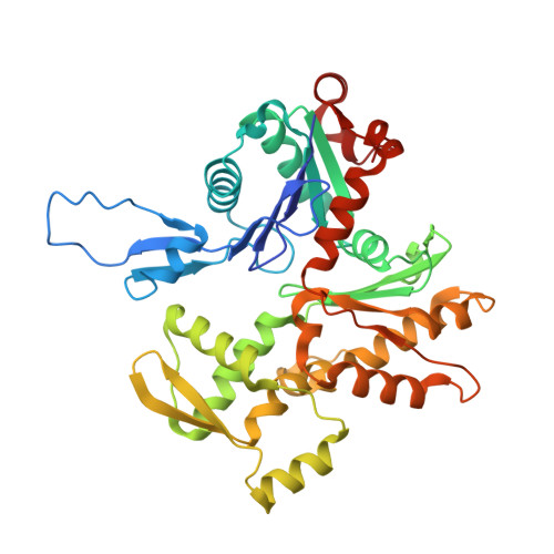
Myosin-7A [auth M]1,935Homo sapiensMutation(s): 0 
Gene Names: MYH7, MYHCB
UniProt & NIH Common Fund Data Resources
Find proteins for P12883 (Homo sapiens)
Explore P12883 
Go to UniProtKB:  P12883
PHAROS:  P12883
GTEx:  ENSG00000092054 
Entity Groups
Sequence Clusters30% Identity50% Identity70% Identity90% Identity95% Identity100% Identity
UniProt GroupP12883
Sequence Annotations
Expand
Reference Sequence
Find similar proteins by:|  3D Structure
Entity ID: 2
MoleculeChains  Sequence LengthOrganismDetailsImage
Actin, alpha cardiac muscle 1
B, C, D, E, F
377Sus scrofaMutation(s): 0 
UniProt
Find proteins for B6VNT8 (Sus scrofa)
Explore B6VNT8 
Go to UniProtKB:  B6VNT8
Entity Groups
Sequence Clusters30% Identity50% Identity70% Identity90% Identity95% Identity100% Identity
UniProt GroupB6VNT8
Sequence Annotations
Expand
Reference Sequence
Find similar proteins by:|  3D Structure
Entity ID: 3
MoleculeChains  Sequence LengthOrganismDetailsImage
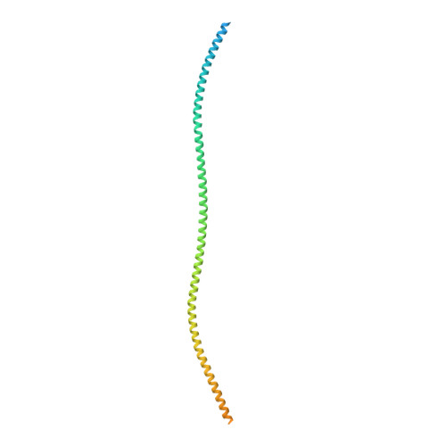
Tropomyosin alpha-1 chainG [auth O],
H [auth P]
284Homo sapiensMutation(s): 0 
Gene Names: TPM1, C15orf13, TMSA
UniProt & NIH Common Fund Data Resources
Find proteins for P09493 (Homo sapiens)
Explore P09493 
Go to UniProtKB:  P09493
PHAROS:  P09493
GTEx:  ENSG00000140416 
Entity Groups
Sequence Clusters30% Identity50% Identity70% Identity90% Identity95% Identity100% Identity
UniProt GroupP09493
Sequence Annotations
Expand
Reference Sequence

Small Molecules

Ligands 2 Unique
IDChains Name / Formula / InChI Key2D Diagram3D Interactions
ADP

Query on ADP


Download:Ideal Coordinates CCD File
I [auth B],
K [auth C],
M [auth D],
O [auth E],
Q [auth F]
ADENOSINE-5'-DIPHOSPHATE
C10 H15 N5 O10 P2
XTWYTFMLZFPYCI-KQYNXXCUSA-N
MG

Query on MG


Download:Ideal Coordinates CCD File
J [auth B],
L [auth C],
N [auth D],
P [auth E],
R [auth F]
MAGNESIUM ION
Mg
JLVVSXFLKOJNIY-UHFFFAOYSA-N

Experimental Data & Validation

Experimental Data

  • Method: ELECTRON MICROSCOPY
  • Resolution: 3.40 Å
  • Aggregation State: FILAMENT 
  • Reconstruction Method: HELICAL 
EM Software:
TaskSoftware PackageVersion
MODEL REFINEMENTPHENIX1.18
RECONSTRUCTIONRELION3.1.1

Structure Validation

View Full Validation Report


Entry History 

& Funding Information

Deposition Data


Funding OrganizationLocationGrant Number
National Institutes of Health/National Heart, Lung, and Blood Institute (NIH/NHLBI)United StatesR01HL036153

Revision History  (Full details and data files)

  • Version 1.0: 2022-11-23
    Type: Initial release
  • Version 1.1: 2022-12-14
    Changes: Database references, Refinement description
  • Version 1.2: 2024-06-19
    Changes: Data collection, Refinement description
  • Version 1.3: 2024-11-13
    Changes: Data collection, Structure summary