8E93

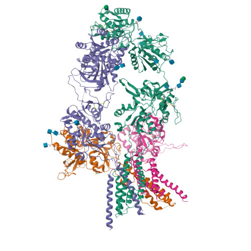
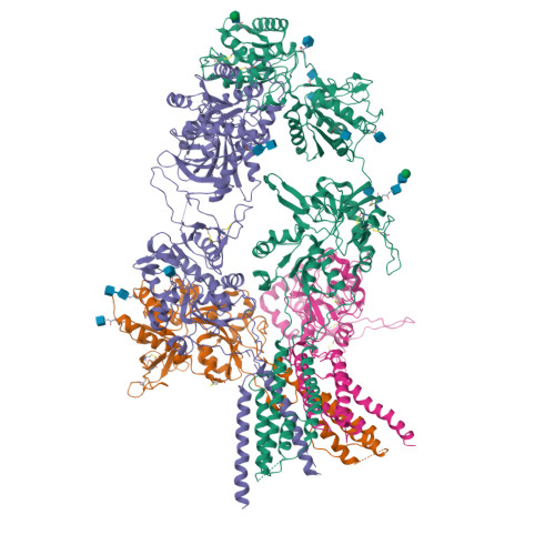
D-cycloserine and glutamate bound Human GluN1a-GluN2C NMDA receptor in splayed conformation

  • Classification: TRANSPORT PROTEIN
  • Organism(s): Homo sapiens
  • Expression System: Spodoptera frugiperda
  • Mutation(s): No 
  • Membrane Protein: Yes  OPMPDBTMmpstruc

  • Deposited: 2022-08-26 Released: 2022-12-07 
  • Deposition Author(s): Chou, T.-H., Furukawa, H.
  • Funding Organization(s): National Institutes of Health/National Institute of Neurological Disorders and Stroke (NIH/NINDS), National Institutes of Health/National Institute of Mental Health (NIH/NIMH)

Experimental Data Snapshot

  • Method: ELECTRON MICROSCOPY
  • Resolution: 3.71 Å
  • Aggregation State: PARTICLE 
  • Reconstruction Method: SINGLE PARTICLE 

wwPDB Validation   3D Report Full Report


This is version 1.2 of the entry. See complete history


Literature

Structural insights into assembly and function of GluN1-2C, GluN1-2A-2C, and GluN1-2D NMDARs.

Chou, T.H.Kang, H.Simorowski, N.Traynelis, S.F.Furukawa, H.

(2022) Mol Cell 82: 4548

  • DOI: https://doi.org/10.1016/j.molcel.2022.10.008
  • Primary Citation of Related Structures:  
    8E92, 8E93, 8E94, 8E96, 8E97, 8E98, 8E99

  • PubMed Abstract: 

    Neurotransmission mediated by diverse subtypes of N-methyl-D-aspartate receptors (NMDARs) is fundamental for basic brain functions and development as well as neuropsychiatric diseases and disorders. NMDARs are glycine- and glutamate-gated ion channels that exist as heterotetramers composed of obligatory GluN1 and GluN2(A-D) and/or GluN3(A-B). The GluN2C and GluN2D subunits form ion channels with distinct properties and spatio-temporal expression patterns. Here, we provide the structures of the agonist-bound human GluN1-2C NMDAR in the presence and absence of the GluN2C-selective positive allosteric potentiator (PAM), PYD-106, the agonist-bound GluN1-2A-2C tri-heteromeric NMDAR, and agonist-bound GluN1-2D NMDARs by single-particle electron cryomicroscopy. Our analysis shows unique inter-subunit and domain arrangements of the GluN2C NMDARs, which contribute to functional regulation and formation of the PAM binding pocket and is distinct from GluN2D NMDARs. Our findings here provide the fundamental blueprint to study GluN2C- and GluN2D-containing NMDARs, which are uniquely involved in neuropsychiatric disorders.


  • Organizational Affiliation

    W.M. Keck Structural Biology Laboratory, Cold Spring Harbor Laboratory, Cold Spring Harbor, NY 11724, USA.


Macromolecules
Find similar proteins by:  (by identity cutoff)  |  3D Structure
Entity ID: 1
MoleculeChains Sequence LengthOrganismDetailsImage
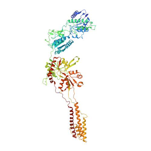
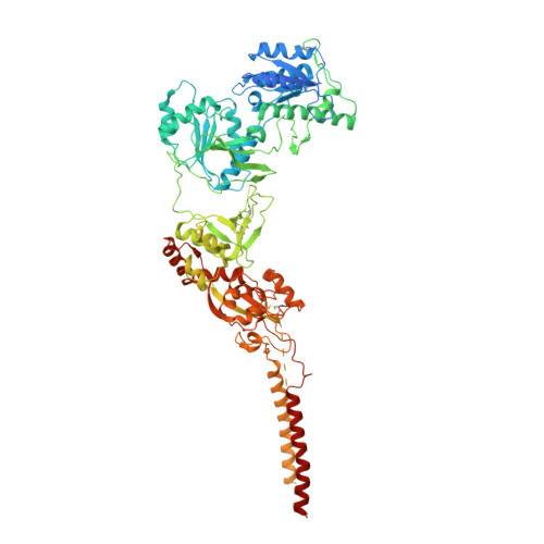
Glutamate receptor ionotropic, NMDA 1
A, C
847Homo sapiensMutation(s): 0 
Gene Names: GRIN1NMDAR1
Membrane Entity: Yes 
UniProt & NIH Common Fund Data Resources
Find proteins for Q05586 (Homo sapiens)
Explore Q05586 
Go to UniProtKB:  Q05586
PHAROS:  Q05586
GTEx:  ENSG00000176884 
Entity Groups  
Sequence Clusters30% Identity50% Identity70% Identity90% Identity95% Identity100% Identity
UniProt GroupQ05586
Glycosylation
Glycosylation Sites: 6Go to GlyGen: Q05586-1
Sequence Annotations
Expand
  • Reference Sequence
Find similar proteins by:  (by identity cutoff)  |  3D Structure
Entity ID: 2
MoleculeChains Sequence LengthOrganismDetailsImage
Glutamate receptor ionotropic, NMDA 2C
B, D
880Homo sapiensMutation(s): 0 
Gene Names: GRIN2CNMDAR2C
Membrane Entity: Yes 
UniProt & NIH Common Fund Data Resources
Find proteins for Q14957 (Homo sapiens)
Explore Q14957 
Go to UniProtKB:  Q14957
PHAROS:  Q14957
GTEx:  ENSG00000161509 
Entity Groups  
Sequence Clusters30% Identity50% Identity70% Identity90% Identity95% Identity100% Identity
UniProt GroupQ14957
Glycosylation
Glycosylation Sites: 2Go to GlyGen: Q14957-1
Sequence Annotations
Expand
  • Reference Sequence
Oligosaccharides

Help

Entity ID: 3
MoleculeChains Length2D Diagram Glycosylation3D Interactions
beta-D-mannopyranose-(1-4)-2-acetamido-2-deoxy-beta-D-glucopyranose-(1-4)-2-acetamido-2-deoxy-beta-D-glucopyranose
E, F
3N-Glycosylation
Glycosylation Resources
GlyTouCan:  G15407YE
GlyCosmos:  G15407YE
GlyGen:  G15407YE
Entity ID: 4
MoleculeChains Length2D Diagram Glycosylation3D Interactions
2-acetamido-2-deoxy-beta-D-glucopyranose-(1-4)-2-acetamido-2-deoxy-beta-D-glucopyranose
G, H
2N-Glycosylation
Glycosylation Resources
GlyTouCan:  G42666HT
GlyCosmos:  G42666HT
GlyGen:  G42666HT
Experimental Data & Validation

Experimental Data

  • Method: ELECTRON MICROSCOPY
  • Resolution: 3.71 Å
  • Aggregation State: PARTICLE 
  • Reconstruction Method: SINGLE PARTICLE 
EM Software:
TaskSoftware PackageVersion
RECONSTRUCTIONcisTEM1.0.2

Structure Validation

View Full Validation Report


Entry History & Funding Information

Deposition Data


Funding OrganizationLocationGrant Number
National Institutes of Health/National Institute of Neurological Disorders and Stroke (NIH/NINDS)United StatesNS111745
National Institutes of Health/National Institute of Mental Health (NIH/NIMH)United StatesMH085926

Revision History  (Full details and data files)

  • Version 1.0: 2022-12-07
    Type: Initial release
  • Version 1.1: 2022-12-14
    Changes: Database references
  • Version 1.2: 2024-10-16
    Changes: Data collection, Structure summary