8E5E

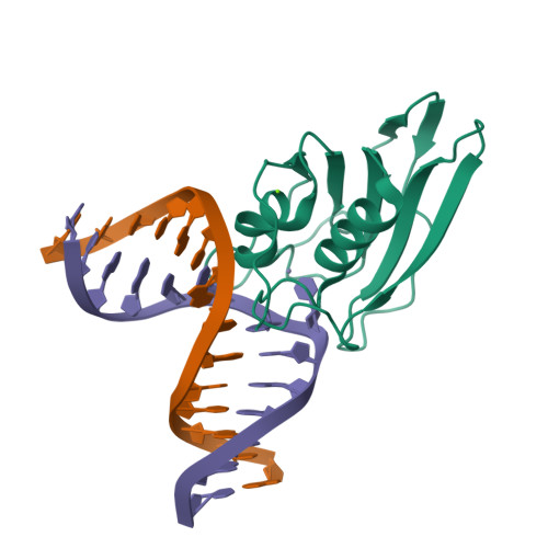
Crystal structure of double-stranded DNA deaminase toxin DddA in complex with DNA with the target cytosine flipped into the active site


Experimental Data Snapshot

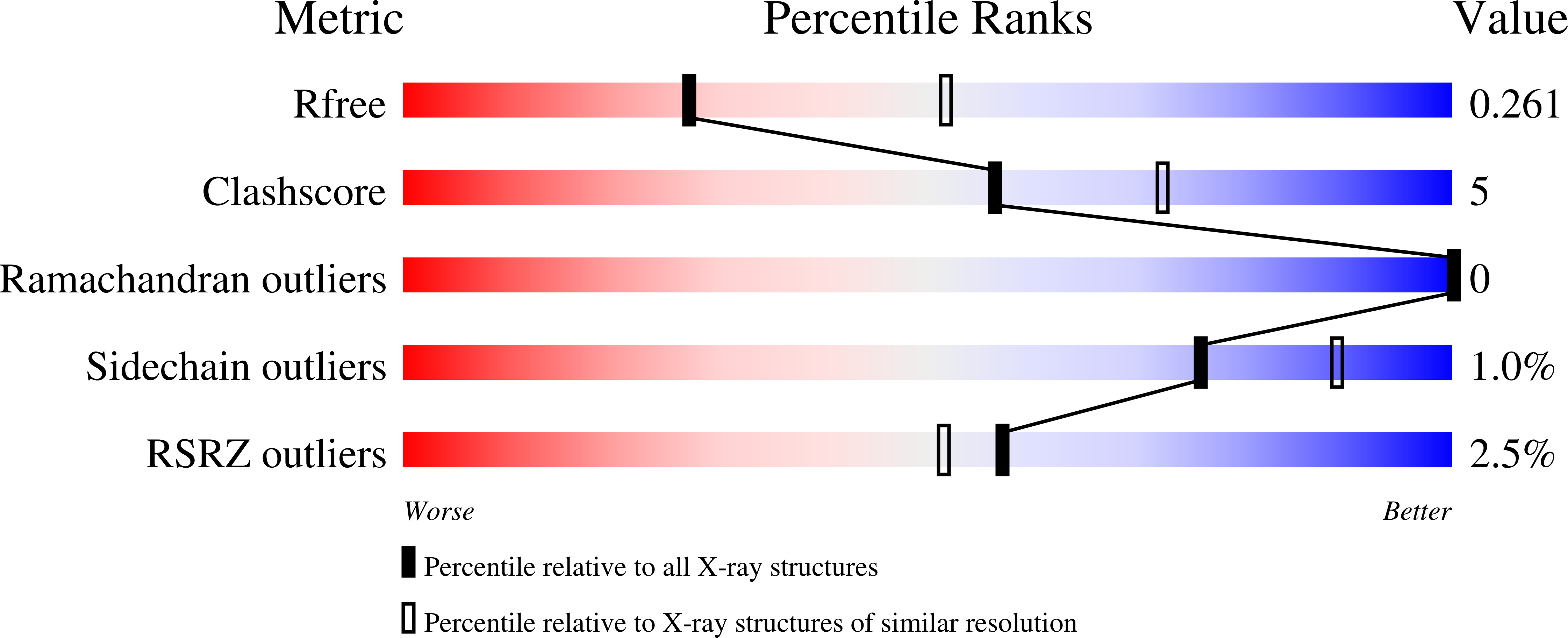
  • Method: X-RAY DIFFRACTION
  • Resolution: 2.62 Å
  • R-Value Free: 0.258 
  • R-Value Work: 0.242 
  • R-Value Observed: 0.243 

Starting Model: experimental
View more details

wwPDB Validation   3D Report Full Report


This is version 1.5 of the entry. See complete history


Literature

Structural basis of sequence-specific cytosine deamination by double-stranded DNA deaminase toxin DddA.

Yin, L.Shi, K.Aihara, H.

(2023) Nat Struct Mol Biol 30: 1153-1159

  • DOI: https://doi.org/10.1038/s41594-023-01034-3
  • Primary Citation of Related Structures:  
    8E5D, 8E5E

  • PubMed Abstract: 

    The interbacterial deaminase toxin DddA catalyzes cytosine-to-uracil conversion in double-stranded (ds) DNA and enables CRISPR-free mitochondrial base editing, but the molecular mechanisms underlying its unique substrate selectivity have remained elusive. Here, we report crystal structures of DddA bound to a dsDNA substrate containing the 5'-TC target motif. These structures show that DddA binds to the minor groove of a sharply bent dsDNA and engages the target cytosine extruded from the double helix. DddA Phe1375 intercalates in dsDNA and displaces the 5' (-1) thymine, which in turn replaces the target (0) cytosine and forms a noncanonical T-G base pair with the juxtaposed guanine. This tandem displacement mechanism allows DddA to locate a target cytosine without flipping it into the active site. Biochemical experiments demonstrate that DNA base mismatches enhance the DddA deaminase activity and relax its sequence selectivity. On the basis of the structural information, we further identified DddA mutants that exhibit attenuated activity or altered substrate preference. Our studies may help design new tools useful in genome editing or other applications.


  • Organizational Affiliation

    Department of Biochemistry, Molecular Biology, and Biophysics, University of Minnesota, Minneapolis, MN, USA.


Macromolecules

Find similar proteins by:  (by identity cutoff)  |  3D Structure
Entity ID: 1
MoleculeChains Sequence LengthOrganismDetailsImage
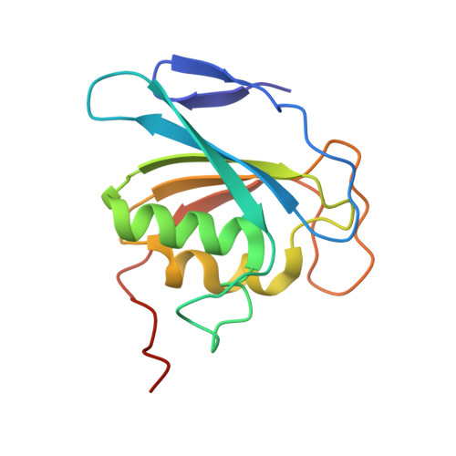
Double-stranded DNA deaminase toxin A139Burkholderia cenocepaciaMutation(s): 1 
Gene Names: dddA
EC: 3.5.4
UniProt
Find proteins for P0DUH5 (Burkholderia cenocepacia (strain H111))
Explore P0DUH5 
Go to UniProtKB:  P0DUH5
Entity Groups  
Sequence Clusters30% Identity50% Identity70% Identity90% Identity95% Identity100% Identity
UniProt GroupP0DUH5
Sequence Annotations
Expand
  • Reference Sequence

Find similar nucleic acids by:  Sequence   |   3D Structure  

Entity ID: 2
MoleculeChains LengthOrganismImage
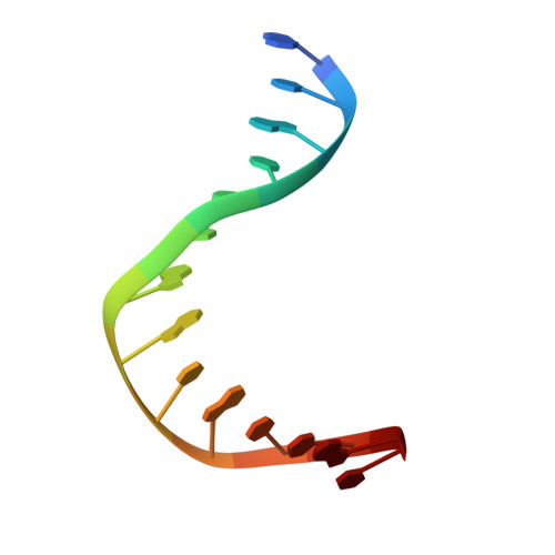
DNA (5'-D(*GP*TP*AP*CP*CP*GP*GP*AP*CP*GP*TP*TP*GP*C)-3')B [auth C]14synthetic construct
Sequence Annotations
Expand
  • Reference Sequence

Find similar nucleic acids by:  Sequence   |   3D Structure  

Entity ID: 3
MoleculeChains LengthOrganismImage
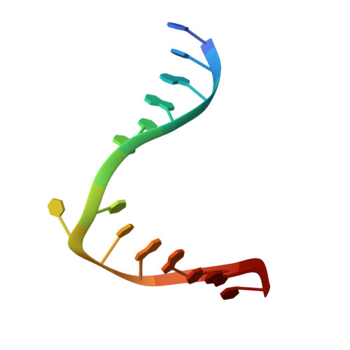
DNA (5'-D(*GP*CP*AP*AP*CP*GP*TP*CP*CP*GP*GP*TP*AP*C)-3')C [auth B]14synthetic construct
Sequence Annotations
Expand
  • Reference Sequence
Experimental Data & Validation

Experimental Data

  • Method: X-RAY DIFFRACTION
  • Resolution: 2.62 Å
  • R-Value Free: 0.258 
  • R-Value Work: 0.242 
  • R-Value Observed: 0.243 
  • Space Group: P 61 2 2
Unit Cell:
Length ( Å )Angle ( ˚ )
a = 62.95α = 90
b = 62.95β = 90
c = 237.078γ = 120
Software Package:
Software NamePurpose
PHENIXrefinement
Aimlessdata scaling
PDB_EXTRACTdata extraction
XDSdata reduction
PHASERphasing

Structure Validation

View Full Validation Report


Entry History & Funding Information

Deposition Data


Funding OrganizationLocationGrant Number
National Institutes of Health/National Cancer Institute (NIH/NCI)United StatesP01-CA234228
National Institutes of Health/National Institute of General Medical Sciences (NIH/NIGMS)United StatesGM118047

Revision History  (Full details and data files)

  • Version 1.0: 2023-05-24
    Type: Initial release
  • Version 1.1: 2023-07-19
    Changes: Database references
  • Version 1.2: 2023-08-02
    Changes: Database references
  • Version 1.3: 2023-08-30
    Changes: Data collection, Database references
  • Version 1.4: 2023-10-25
    Changes: Refinement description
  • Version 1.5: 2024-09-18
    Changes: Structure summary