8E0V | pdb_00008e0v

DAHP (3-deoxy-D-arabinoheptulosonate-7-phosphate) Synthase complexed with Mn(II), PEP, and Pi in unbound:(bound)2:other Conformations


Experimental Data Snapshot

  • Method: X-RAY DIFFRACTION
  • Resolution: 2.30 Å
  • R-Value Free: 
    0.228 (Depositor), 0.227 (DCC) 
  • R-Value Work: 
    0.194 (Depositor), 0.194 (DCC) 
  • R-Value Observed: 
    0.195 (Depositor) 

Starting Model: experimental
View more details

wwPDB Validation 3D Report Full Report

Validation slider image for 8E0V

Ligand Structure Quality Assessment 


This is version 1.2 of the entry. See complete history

Literature

Role of Half-of-Sites Reactivity and Inter-Subunit Communications in DAHP Synthase Catalysis and Regulation.

Balachandran, N.Grainger, R.A.Rob, T.Liuni, P.Wilson, D.J.Junop, M.S.Berti, P.J.

(2022) Biochemistry 61: 2229-2240

  • DOI: https://doi.org/10.1021/acs.biochem.2c00465
  • Primary Citation Related Structures: 
    8E0S, 8E0T, 8E0U, 8E0V, 8E0X, 8E0Y, 8E0Z

  • PubMed Abstract: 

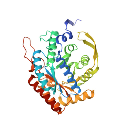
    α-Carboxyketose synthases, including 3-deoxy-d- arabino heptulosonate 7-phosphate synthase (DAHPS), are long-standing targets for inhibition. They are challenging targets to create tight-binding inhibitors against, and inhibitors often display half-of-sites binding and partial inhibition. Half-of-sites inhibition demonstrates the existence of inter-subunit communication in DAHPS. We used X-ray crystallography and spatially resolved hydrogen-deuterium exchange (HDX) to reveal the structural and dynamic bases for inter-subunit communication in Escherichia coli DAHPS(Phe), the isozyme that is feedback-inhibited by phenylalanine. Crystal structures of this homotetrameric (dimer-of-dimers) enzyme are invariant over 91% of its sequence. Three variable loops make up 8% of the sequence and are all involved in inter-subunit contacts across the tight-dimer interface. The structures have pseudo-twofold symmetry indicative of inter-subunit communication across the loose-dimer interface, with the diagonal subunits B and C always having the same conformation as each other, while subunits A and D are variable. Spatially resolved HDX reveals contrasting responses to ligand binding, which, in turn, affect binding of the second substrate, erythrose-4-phosphate (E4P). The N -terminal peptide, M1-E12, and the active site loop that binds E4P, F95-K105, are key parts of the communication network. Inter-subunit communication appears to have a catalytic role in all α-carboxyketose synthase families and a regulatory role in some members.


  • Organizational Affiliation
    • Department of Biochemistry, Molecular Biology Lab, Western University, London, Ontario N6A 5C1, Canada.

Macromolecule Content 

  • Total Structure Weight: 153.75 kDa 
  • Atom Count: 11,229 
  • Modeled Residue Count: 1,371 
  • Deposited Residue Count: 1,404 
  • Unique protein chains: 1

Macromolecules

Find similar proteins by:|  3D Structure
Entity ID: 1
MoleculeChains  Sequence LengthOrganismDetailsImage
Phospho-2-dehydro-3-deoxyheptonate aldolase, Phe-sensitive
A, B, C, D
351Escherichia coli K-12Mutation(s): 0 
Gene Names: aroGb0754JW0737
EC: 2.5.1.54
UniProt
Find proteins for P0AB91 (Escherichia coli (strain K12))
Explore P0AB91 
Go to UniProtKB:  P0AB91
Entity Groups
Sequence Clusters30% Identity50% Identity70% Identity90% Identity95% Identity100% Identity
UniProt GroupP0AB91
Sequence Annotations
Expand
Reference Sequence

Small Molecules

Ligands 4 Unique
IDChains Name / Formula / InChI Key2D Diagram3D Interactions
GAL

Query on GAL


Download:Ideal Coordinates CCD File
F [auth A],
G [auth A],
N [auth C]
beta-D-galactopyranose
C6 H12 O6
WQZGKKKJIJFFOK-FPRJBGLDSA-N
PEP
(Subject of Investigation/LOI)

Query on PEP


Download:Ideal Coordinates CCD File
I [auth B],
L [auth C]
PHOSPHOENOLPYRUVATE
C3 H5 O6 P
DTBNBXWJWCWCIK-UHFFFAOYSA-N
PO4
(Subject of Investigation/LOI)

Query on PO4


Download:Ideal Coordinates CCD File
J [auth B],
M [auth C]
PHOSPHATE ION
O4 P
NBIIXXVUZAFLBC-UHFFFAOYSA-K
MN
(Subject of Investigation/LOI)

Query on MN


Download:Ideal Coordinates CCD File
E [auth A],
H [auth B],
K [auth C],
O [auth D]
MANGANESE (II) ION
Mn
WAEMQWOKJMHJLA-UHFFFAOYSA-N

Experimental Data & Validation

Experimental Data

  • Method: X-RAY DIFFRACTION
  • Resolution: 2.30 Å
  • R-Value Free:  0.228 (Depositor), 0.227 (DCC) 
  • R-Value Work:  0.194 (Depositor), 0.194 (DCC) 
  • R-Value Observed: 0.195 (Depositor) 
Space Group: C 1 2 1
Unit Cell:
Length ( Å )Angle ( ˚ )
a = 210.193α = 90
b = 52.908β = 115.711
c = 150.481γ = 90
Software Package:
Software NamePurpose
PHENIXrefinement
autoPROCdata reduction
autoPROCdata scaling
PHASERphasing

Structure Validation

View Full Validation Report


Ligand Structure Quality Assessment 


Entry History 

& Funding Information

Deposition Data


Funding OrganizationLocationGrant Number
Natural Sciences and Engineering Research Council (NSERC, Canada)Canada2017-06712
Natural Sciences and Engineering Research Council (NSERC, Canada)Canada262034-2013
Natural Sciences and Engineering Research Council (NSERC, Canada)CanadaRGPIN 480432
Canadian Institutes of Health Research (CIHR)CanadaMOP-64422
Canadian Institutes of Health Research (CIHR)CanadaMOP-166070

Revision History  (Full details and data files)

  • Version 1.0: 2022-10-12
    Type: Initial release
  • Version 1.1: 2022-11-02
    Changes: Database references
  • Version 1.2: 2023-10-18
    Changes: Data collection, Refinement description