8DZH | pdb_00008dzh

Structure of SARS-CoV-2 Omicron BA.1.1.529 Spike trimer with two RBDs down in complex with the Fab fragment of human neutralizing antibody MB.02


Experimental Data Snapshot

  • Method: ELECTRON MICROSCOPY
  • Resolution: 3.20 Å
  • Aggregation State: PARTICLE 
  • Reconstruction Method: SINGLE PARTICLE 

wwPDB Validation 3D Report Full Report

Validation slider image for 8DZH

This is version 1.4 of the entry. See complete history

Literature

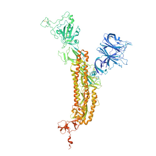
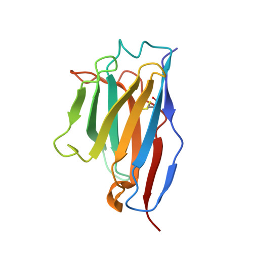
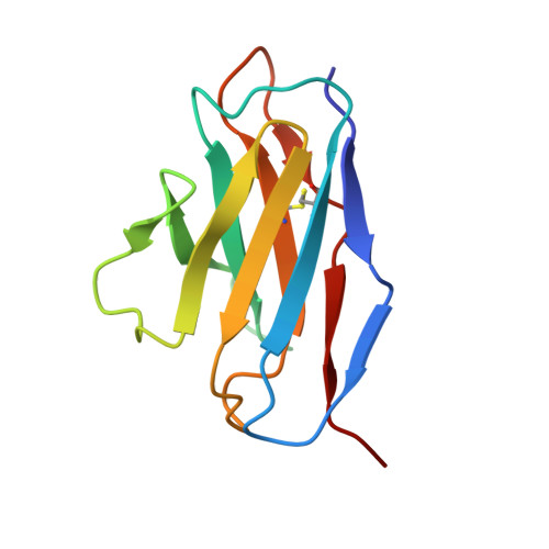
Function and Cryo-EM structures of broadly potent bispecific antibodies against multiple SARS-CoV-2 Omicron sublineages.

Ren, P.Hu, Y.Peng, L.Yang, L.Suzuki, K.Fang, Z.Bai, M.Zhou, L.Feng, Y.Zou, Y.Xiong, Y.Chen, S.

(2023) Signal Transduct Target Ther 8: 281-281

  • DOI: https://doi.org/10.1038/s41392-023-01509-1
  • Primary Citation Related Structures: 
    8DZH, 8DZI

  • Organizational Affiliation
    • Department of Genetics, Yale University School of Medicine, New Haven, CT, USA.

Macromolecule Content 

  • Total Structure Weight: 517.39 kDa 
  • Atom Count: 31,956 
  • Modeled Residue Count: 3,966 
  • Deposited Residue Count: 4,536 
  • Unique protein chains: 3

Macromolecules

Find similar proteins by:|  3D Structure
Entity ID: 1
MoleculeChains  Sequence LengthOrganismDetailsImage
Spike glycoproteinA [auth B],
B [auth A],
C
1,285Severe acute respiratory syndrome coronavirus 2Mutation(s): 10 
Gene Names: S2
UniProt
Find proteins for P0DTC2 (Severe acute respiratory syndrome coronavirus 2)
Explore P0DTC2 
Go to UniProtKB:  P0DTC2
Entity Groups
Sequence Clusters30% Identity50% Identity70% Identity90% Identity95% Identity100% Identity
UniProt GroupP0DTC2
Glycosylation
Glycosylation Sites: 16Go to GlyGen: P0DTC2-1
Sequence Annotations
Expand
Reference Sequence
Find similar proteins by:|  3D Structure
Entity ID: 2
MoleculeChains  Sequence LengthOrganismDetailsImage
antibody MB.02 heavy chainD [auth E],
F [auth H],
H [auth I]
118Homo sapiensMutation(s): 0 
Find similar proteins by:|  3D Structure
Entity ID: 3
MoleculeChains  Sequence LengthOrganismDetailsImage
antibody MB.02 light chainE [auth D],
G [auth L],
I [auth J]
109Homo sapiensMutation(s): 0 

Oligosaccharides

Help  

Small Molecules

Ligands 1 Unique
IDChains Name / Formula / InChI Key2D Diagram3D Interactions
NAG
(Subject of Investigation/LOI)

Query on NAG


Download:Ideal Coordinates CCD File
AB [auth C]
BB [auth C]
CB [auth C]
KA [auth B]
LA [auth B]
AB [auth C],
BB [auth C],
CB [auth C],
KA [auth B],
LA [auth B],
MA [auth B],
NA [auth B],
OA [auth B],
PA [auth B],
QA [auth B],
RA [auth A],
SA [auth A],
TA [auth A],
UA [auth A],
VA [auth A],
WA [auth A],
XA [auth C],
YA [auth C],
ZA [auth C]
2-acetamido-2-deoxy-beta-D-glucopyranose
C8 H15 N O6
OVRNDRQMDRJTHS-FMDGEEDCSA-N

Experimental Data & Validation

Experimental Data

  • Method: ELECTRON MICROSCOPY
  • Resolution: 3.20 Å
  • Aggregation State: PARTICLE 
  • Reconstruction Method: SINGLE PARTICLE 
EM Software:
TaskSoftware PackageVersion
MODEL REFINEMENTPHENIX

Structure Validation

View Full Validation Report


Entry History 

& Funding Information

Deposition Data


Funding OrganizationLocationGrant Number
National Institutes of Health/National Cancer Institute (NIH/NCI)United States--

Revision History  (Full details and data files)

  • Version 1.0: 2022-08-31
    Type: Initial release
  • Version 1.1: 2023-03-29
    Changes: Database references
  • Version 1.2: 2023-11-15
    Changes: Data collection, Database references
  • Version 1.3: 2024-10-30
    Changes: Data collection, Structure summary
  • Version 1.4: 2025-05-21
    Changes: Data collection