8DP3 | pdb_00008dp3

Crystal structure of coxsackievirus B3 cloverleaf RNA replication element


Experimental Data Snapshot

  • Method: X-RAY DIFFRACTION
  • Resolution: 1.91 Å
  • R-Value Free: 
    0.259 (Depositor), 0.261 (DCC) 
  • R-Value Work: 
    0.209 (Depositor), 0.209 (DCC) 
  • R-Value Observed: 
    0.211 (Depositor) 

Starting Model: experimental
View more details

wwPDB Validation 3D Report Full Report

Validation slider image for 8DP3

Ligand Structure Quality Assessment 


This is version 1.2 of the entry. See complete history

Literature

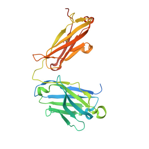
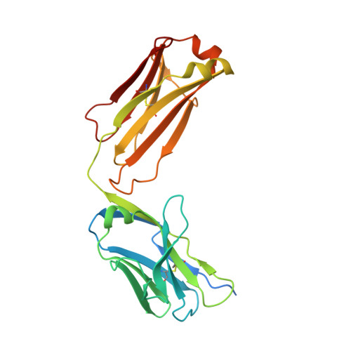
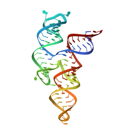
Crystal structure of a highly conserved enteroviral 5' cloverleaf RNA replication element.

Das, N.K.Hollmann, N.M.Vogt, J.Sevdalis, S.E.Banna, H.A.Ojha, M.Koirala, D.

(2023) Nat Commun 14: 1955-1955

  • DOI: https://doi.org/10.1038/s41467-023-37658-8
  • Primary Citation Related Structures: 
    8DP3

  • PubMed Abstract: 

    The extreme 5'-end of the enterovirus RNA genome contains a conserved cloverleaf-like domain that recruits 3CD and PCBP proteins required for initiating genome replication. Here, we report the crystal structure at 1.9 Å resolution of this domain from the CVB3 genome in complex with an antibody chaperone. The RNA folds into an antiparallel H-type four-way junction comprising four subdomains with co-axially stacked sA-sD and sB-sC helices. Long-range interactions between a conserved A40 in the sC-loop and Py-Py helix within the sD subdomain organize near-parallel orientations of the sA-sB and sC-sD helices. Our NMR studies confirm that these long-range interactions occur in solution and without the chaperone. The phylogenetic analyses indicate that our crystal structure represents a conserved architecture of enteroviral cloverleaf-like domains, including the A40 and Py-Py interactions. The protein binding studies further suggest that the H-shape architecture provides a ready-made platform to recruit 3CD and PCBP2 for viral replication.


  • Organizational Affiliation
    • Department of Chemistry and Biochemistry, University of Maryland Baltimore County, Baltimore, MD, 21250, USA.

Macromolecule Content 

  • Total Structure Weight: 82.09 kDa 
  • Atom Count: 5,657 
  • Modeled Residue Count: 531 
  • Deposited Residue Count: 584 
  • Unique protein chains: 2
  • Unique nucleic acid chains: 1

Macromolecules


Find similar proteins by:|  3D Structure
Entity ID: 1
MoleculeChains  Sequence LengthOrganismDetailsImage
Fab BL3-6 Heavy ChainA [auth H]256synthetic constructMutation(s): 0 
Find similar proteins by:|  3D Structure
Entity ID: 2
MoleculeChains  Sequence LengthOrganismDetailsImage
Fab BL3-6 Light ChainB [auth L]238synthetic constructMutation(s): 0 
Find similar nucleic acids by:  (by identity cutoff) 
Entity ID: 3
MoleculeChains LengthOrganismImage
RNA (90-MER)C [auth R]90Coxsackievirus B3
Sequence Annotations
Expand
Reference Sequence

Small Molecules

Ligands 1 Unique
IDChains Name / Formula / InChI Key2D Diagram3D Interactions
PO4
(Subject of Investigation/LOI)

Query on PO4


Download:Ideal Coordinates CCD File
D [auth L]PHOSPHATE ION
O4 P
NBIIXXVUZAFLBC-UHFFFAOYSA-K

Experimental Data & Validation

Experimental Data

  • Method: X-RAY DIFFRACTION
  • Resolution: 1.91 Å
  • R-Value Free:  0.259 (Depositor), 0.261 (DCC) 
  • R-Value Work:  0.209 (Depositor), 0.209 (DCC) 
  • R-Value Observed: 0.211 (Depositor) 
Space Group: C 1 2 1
Unit Cell:
Length ( Å )Angle ( ˚ )
a = 122.173α = 90
b = 48.698β = 112.92
c = 144.653γ = 90
Software Package:
Software NamePurpose
PHENIXrefinement
PDB_EXTRACTdata extraction
XDSdata reduction
Aimlessdata scaling
PHENIXphasing

Structure Validation

View Full Validation Report


Ligand Structure Quality Assessment 


Entry History 

& Funding Information

Deposition Data


Funding OrganizationLocationGrant Number
Other governmentUnited StatesStart-up

Revision History  (Full details and data files)

  • Version 1.0: 2023-04-19
    Type: Initial release
  • Version 1.1: 2023-10-25
    Changes: Data collection, Refinement description
  • Version 1.2: 2024-10-30
    Changes: Structure summary