8DI5

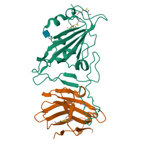
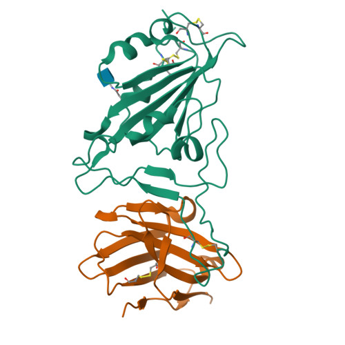
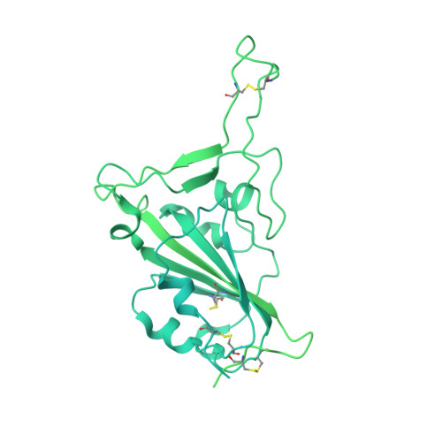
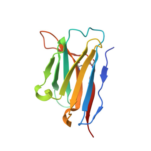
Cryo-EM structure of SARS-CoV-2 Beta (B.1.351) spike protein in complex with VH domain F6 (focused refinement of RBD and VH F6)


Experimental Data Snapshot

  • Method: ELECTRON MICROSCOPY
  • Resolution: 3.04 Å
  • Aggregation State: PARTICLE 
  • Reconstruction Method: SINGLE PARTICLE 

wwPDB Validation   3D Report Full Report


This is version 1.1 of the entry. See complete history


Literature

Potent and broad neutralization of SARS-CoV-2 variants of concern (VOCs) including omicron sub-lineages BA.1 and BA.2 by biparatopic human VH domains.

Chen, C.Saville, J.W.Marti, M.M.Schafer, A.Cheng, M.H.Mannar, D.Zhu, X.Berezuk, A.M.Banerjee, A.Sobolewski, M.D.Kim, A.Treat, B.R.Da Silva Castanha, P.M.Enick, N.McCormick, K.D.Liu, X.Adams, C.Hines, M.G.Sun, Z.Chen, W.Jacobs, J.L.Barratt-Boyes, S.M.Mellors, J.W.Baric, R.S.Bahar, I.Dimitrov, D.S.Subramaniam, S.Martinez, D.R.Li, W.

(2022) iScience 25: 104798-104798

  • DOI: https://doi.org/10.1016/j.isci.2022.104798
  • Primary Citation of Related Structures:  
    8DI5

  • PubMed Abstract: 

    The emergence of SARS-CoV-2 variants of concern (VOCs) requires the development of next-generation biologics with high neutralization breadth. Here, we characterized a human V H domain, F6, which we generated by sequentially panning large phage-displayed V H libraries against receptor binding domains (RBDs) containing VOC mutations. Cryo-EM analyses reveal that F6 has a unique binding mode that spans a broad surface of the RBD and involves the antibody framework region. Attachment of an Fc region to a fusion of F6 and ab8, a previously characterized V H domain, resulted in a construct (F6-ab8-Fc) that broadly and potently neutralized VOCs including Omicron. Additionally, prophylactic treatment using F6-ab8-Fc reduced live Beta (B.1.351) variant viral titers in the lungs of a mouse model. Our results provide a new potential therapeutic against SARS-CoV-2 variants including Omicron and highlight a vulnerable epitope within the spike that may be exploited to achieve broad protection against circulating variants.


  • Organizational Affiliation

    Center for Antibody Therapeutics, Division of Infectious Diseases, Department of Medicine, University of Pittsburgh Medical School, Pittsburgh, PA, USA.


Macromolecules
Find similar proteins by:  (by identity cutoff)  |  3D Structure
Entity ID: 1
MoleculeChains Sequence LengthOrganismDetailsImage
Spike glycoproteinA [auth C]1,288Severe acute respiratory syndrome coronavirus 2Mutation(s): 18 
Gene Names: S2
UniProt
Find proteins for P0DTC2 (Severe acute respiratory syndrome coronavirus 2)
Explore P0DTC2 
Go to UniProtKB:  P0DTC2
Entity Groups  
Sequence Clusters30% Identity50% Identity70% Identity90% Identity95% Identity100% Identity
UniProt GroupP0DTC2
Glycosylation
Glycosylation Sites: 1Go to GlyGen: P0DTC2-1
Sequence Annotations
Expand
  • Reference Sequence
Find similar proteins by:  (by identity cutoff)  |  3D Structure
Entity ID: 2
MoleculeChains Sequence LengthOrganismDetailsImage
VH F6B [auth H]118Homo sapiensMutation(s): 0 
Entity Groups  
Sequence Clusters30% Identity50% Identity70% Identity90% Identity95% Identity100% Identity
Sequence Annotations
Expand
  • Reference Sequence
Small Molecules
Ligands 1 Unique
IDChains Name / Formula / InChI Key2D Diagram3D Interactions
NAG
Query on NAG

Download Ideal Coordinates CCD File 
C
2-acetamido-2-deoxy-beta-D-glucopyranose
C8 H15 N O6
OVRNDRQMDRJTHS-FMDGEEDCSA-N
Experimental Data & Validation

Experimental Data

  • Method: ELECTRON MICROSCOPY
  • Resolution: 3.04 Å
  • Aggregation State: PARTICLE 
  • Reconstruction Method: SINGLE PARTICLE 

Structure Validation

View Full Validation Report


Entry History & Funding Information

Deposition Data


Funding OrganizationLocationGrant Number
Canada Excellence Research Chair AwardCanadaPrecision Cancer Drug Design
Other governmentCanadaCOVID-19 research

Revision History  (Full details and data files)

  • Version 1.0: 2022-08-24
    Type: Initial release
  • Version 1.1: 2024-10-09
    Changes: Data collection, Structure summary